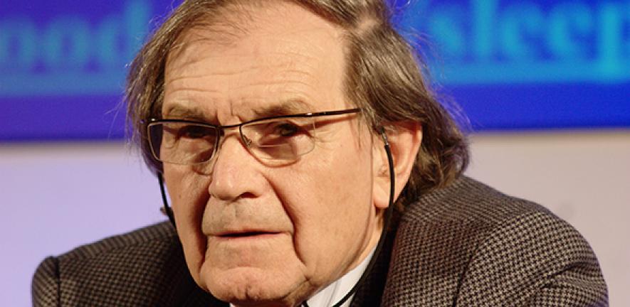
Professor Chris Abell FRS, FMedSci (1957 – 2020)

source: cam.ac.uk
The University is saddened to announce that Professor Chris Abell, Pro-Vice-Chancellor for Research, Professor of Biological Chemistry and Todd-Hamied Fellow of Christ’s College, has died suddenly at the age of 62.
A biological chemist, he was a pioneer in the field of fragment-based drug discovery, a successful entrepreneur, a founding director of Cambridge Enterprise, and the University’s first Director of Postdoctoral Affairs.
A major focus of his highly interdisciplinary research in the Department of Chemistry was to understand the mechanisms of key enzymes and develop approaches to their inhibition, an approach that could lead to new treatments for diseases such as tuberculosis, cystic fibrosis and cancer.
The advances he made in fragment-based drug discovery led him to co-found Astex, a world-leading company in this area, in 1999. Fragment-based approaches are now adopted throughout the pharmaceutical industry and in many academic laboratories.
He also made major contributions to the development of microfluidic microdroplets as a platform for experimental science, with applications in cell biology, chemistry and materials science. This interest resulted in the co-founding of Sphere Fluidics (2010) and Aqdot (2013).
He was an undergraduate and postgraduate student at St John’s College, Cambridge, before conducting postdoctoral research at Brown University, USA. He was named a Fellow of the Academy of Medical Sciences in 2012 and a Fellow of the Royal Society in 2016.
Vice-Chancellor Professor Stephen J Toope said: “Chris’ death is a huge loss to the University, and to me personally. Our thoughts and our deepest sympathies are with his wife, Dr Katherine Abell, their son Daniel, and with Chris’ friends and colleagues at the Department of Chemistry, at the Research Operations and Research Strategy Offices, and at Christ’s College.”
Professor Jane Stapleton, Master of Christ’s College, said: “In Christ’s we are devastated by the shocking news of the death of Chris Abell, our warm, wise friend. He has long been held in the greatest esteem by the College to which he devoted so much of his remarkable energy.”
Dr James Keeler, Head of the Department of Chemistry, said: “Chris has for many years been a leading figure in the field of biological chemistry and has been responsible for significant advances in the field. He has also been conspicuously successful in commercialising aspects of his work, most notably as co-founder of Astex. Chris is remembered by us all as an outstanding scientist, a valued and loyal colleague, and a tireless champion for the Department and the University.”
A digital condolences book has been set up at: www.remembr.com/professor.chris.abell.

The text in this work is licensed under a Creative Commons Attribution 4.0 International License. Images, including our videos, are Copyright ©University of Cambridge and licensors/contributors as identified. All rights reserved. We make our image and video content available in a number of ways – as here, on our main website under its Terms and conditions, and on a range of channels including social media that permit your use and sharing of our content under their respective Terms.