All posts by Admin

Scientists Supercharge Shellfish To Tackle Vitamin Deficiency in Humans

Oysters
source: www.cam.ac.uk

Cambridge scientists have developed a new way to fortify shellfish to tackle human nutrient deficiencies which cause severe health problems across the world. The team is now working with major seafood manufacturers to further test their microencapsulation technology, or “Vitamin Bullets”.

Targeted use of this technology in regions worst affected by nutrient deficiencies … could help improve the health of millions

David Willer

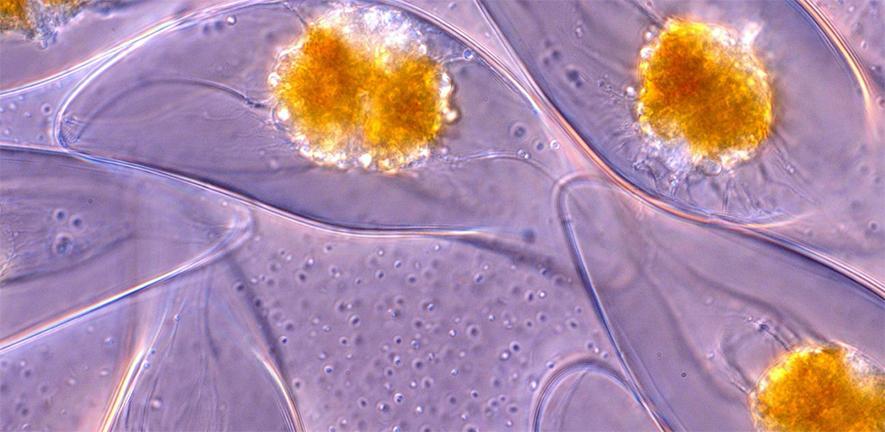
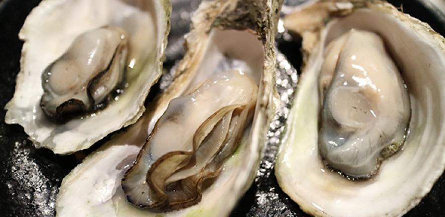
Over two billion people worldwide are nutrient deficient, leading to a wide range of serious health problems. Fortifying food with micronutrients is already an industry standard for enhancing public health but now scientists at Cambridge’s Department of Zoology have teamed up with Cambridge-based company BioBullets to supercharge one of the world’s most healthy and sustainable sources of animal protein: bivalve shellfish such as oysters, clams and mussels.

Dr David Aldridge and PhD student David Willer have produced the world’s first microcapsule specially designed to deliver nutrients to bivalves which are beneficial to human health. These “Vitamin Bullets” – manufactured under patent by Aldridge’s company, BioBullets – are tailored for optimal size, shape, buoyancy and to appeal to shellfish.

This breakthrough, described in a study published today in the journal Frontiers in Nutrition, is particularly valuable because when we eat bivalves, we consume the entire organism including its gut, meaning that we digest the nutrients which the animals consumed towards the end of their lives. This makes bivalve shellfish the ideal target for nutritional fortification.

In their Cambridge laboratory, the scientists trialled Vitamin A and D fortified microcapsules on over 100 oysters to identify the optimal dose. They also established that this should be fed for 8 hours towards the end of “depuration”, the period in which bivalves are held in cleansing tanks after being harvested.

The team found that fortified oysters delivered around 100 times more Vitamin A, and over 150 times more Vitamin D, than natural oysters. Even more importantly, they dramatically outperformed salmon, one of the best natural sources of these vitamins. The fortified oysters provided more than 26 times more Vitamin A and over four times more Vitamin D than salmon. The scientists found that a serving of just two of their supercharged shellfish provided enough Vitamin A and D to meet human Recommended Dietary Allowance (RDAs).

Vitamin A and D deficiencies pose a particularly serious public health challenge – in Ghana more than 76% of children are Vitamin A deficient, causing widespread mortality and blindness. In India, 85% of the population is Vitamin D deficient, which causes cardiovascular diseases, osteoporosis, and rickets. Even in the US, over 40% of people are Vitamin D deficient.

David Willer said: “We have demonstrated a cheap and effective way to get micronutrients into a sustainable and delicious source of protein. Targeted use of this technology in regions worst affected by nutrient deficiencies, using carefully selected bivalve species and micronutrients, could help improve the health of millions, while also reducing the harm that meat production is doing to the environment”.

David Aldridge said: “We are very excited about BioBullets’ potential. We are now establishing links with some of the world’s biggest seafood manufacturers to drive a step change in the sustainability and nutritional value of the seafood that we consume.”

Bivalves have a higher protein content than beef, are a rich source of omega-3 fatty acids, and have some of the highest levels of key minerals of all animal foods. Nevertheless, the nutrients that they deliver naturally is unlikely to solve global deficiencies. These shellfish are also highly sustainable to farm, having a far lower environmental footprint than animal meat or fish, and lower even than many plant crops such as wheat, soya, and rice.

Bivalves are a highly affordable food source when produced at large scale and the global market is rapidly expanding. Production in China alone has grown 1000-fold since 1980 and there is great potential to sustainably expand bivalve aquaculture worldwide, with over 1,500,000 km2 available for sustainable low-cost industry development, particularly around the west coast of Africa and India.

The researchers point out that consumers in poorer regions where vitamin deficiencies are most prevalent are more likely to buy slightly more expensive fortified food than to make additional purchases to take supplement pills. They calculate that fortification adds just $0.0056 to the cost of producing a single oyster.

David Willer is supported by the Biotechnology and Biological Sciences Research Council; and David Aldridge is supported by The University of Cambridge, St Catharine’s College and Corpus Christi College.
Reference:

D.F. Willer & D.C. Aldridge, ‘Vitamin bullets. Microencapsulated feeds to fortify shellfish and tackle human nutrient deficiencies’, Frontiers in Nutrition (July 2020). DOI: 10.3389/fnut.2020.00102


Read on

Learn more about the work of David Willer and Dr David Aldridge in this feature.


Creative Commons License
The text in this work is licensed under a Creative Commons Attribution 4.0 International License. Images, including our videos, are Copyright ©University of Cambridge and licensors/contributors as identified.  All rights reserved. We make our image and video content available in a number of ways – as here, on our main website under its Terms and conditions, and on a range of channels including social media that permit your use and sharing of our content under their respective Terms.

Mixed Early Progress Highlights Need For Sustained Support For Pupils With English As An Additional Language

source: www.cam.ac.uk

Newly-arrived pupils who speak English as an additional language (EAL) often make ‘mixed’ linguistic and academic progress during their first years in British schools, which need a proper framework to give them sustained support, a study suggests.

At the moment, it is often left to individual teachers or schools to decide how to handle the challenges of a multilingual classroom.

Karen Forbes

The finding is one of numerous results and recommendations in a new book about the language development of EAL pupils, and its impact on their attainment and social integration. The book, authored by a team of academics from Cambridge, Anglia Ruskin and Durham Universities, examines the complex relationship between language, education and the social integration of newcomer migrant EAL students.

According to the School Census, there are currently over 1.5 million EAL pupils in England, and the proportion is steadily rising. The trend is similar in many other English-speaking countries.

The book builds on three years of research involving over 40 schools across the East of England, funded by the Bell Foundation, and highlights much good practice by teachers working in multilingual classrooms. But it also points to inconsistencies and gaps in support for EAL pupils, stemming from an absence of national guidelines, targeted assessment, and systemic problems in areas such as teacher training and school-parent communication.

EAL pupils themselves were found to make uneven progress during their first two years in English schools. While many became competent English-speakers, their written English frequently lagged behind. The authors suggest this pattern may be further exacerbated by reductions in funding for EAL support.

As well as analysing the progress of EAL pupils, the study proposes a model for a more inclusive approach to teaching EAL students.

Dr Karen Forbes, Lecturer at the Faculty of Education, University of Cambridge, said: “At the moment, it is often left to individual teachers or schools to decide how to handle the challenges of a multilingual classroom. While many do excellent work, EAL pupils inevitably have a variable experience. Teachers and schools should be able to draw on a structured framework and a proper knowledge base so that they can give these pupils the sustained linguistic and educational support they often need.”

The research suggests that while many schools rightly prioritise the integration of EAL learners into mainstream lessons, some will need ongoing, one-to-one support, especially with developing more academic English, long past the point where they appear socially-integrated and able to hold a casual conversation.

This is just one symptom of a wider need to provide schools with a structural basis to give EAL learners individualised, ‘child-centred’ support, the authors argue. They stress that the ‘EAL’ label does not describe one type of pupil, but encompasses a wide range of previous educational experiences, interests and skills.

Encouragingly, many of the schools surveyed actively encouraged an inclusive and positive environment for EAL pupils. Teachers also employed various tactics that could form part of a wider framework to support them, such as group learning and buddy systems, translated texts and different visual aids.

But the study finds that many such interventions are devised locally, by schools or individual teachers, absent more structured or systematic guidance. This can lead to inconsistencies: for example, teachers varied their approach to when EAL pupils could use their home language, which often left students confused about when to use English.

The researchers argue that other mechanisms are needed to give teachers a more solid foundation for working with EAL pupils. Teachers consistently enthused, for example, about the ‘vital’ support provided by dedicated EAL co-ordinators and bilingual support staff. But many schools that the researchers surveyed have struggled to sustain such services given that funding is no longer ring-fenced for this purpose.

The book also highlights the need for more EAL-specific, specialist training for teachers, both for their professional practice and to help them work successfully with local minority-ethnic and migrant communities, especially those unfamiliar with the English system of education. This is only covered briefly in most teacher-training courses, and rarely forms part of their continuing professional development or ‘on the job’ learning.

Critically, the researchers also suggest that parents of EAL pupils and their communities are an untapped resource of knowledge, strong educational values and expertise.

The researchers found that many parents of EAL children have a high level of interest in their children’s education, but often are not sufficiently supported to understand context-specific curriculum choices, modes of assessment or school expectations. They argue that, as well as providing translated information and induction materials, schools should establish mechanisms such as EAL parents’ networks, empowering parents within school governance structures to inform the way that they support migrant pupils, ensure that they achieve their potential, and promote positive experiences in school.

“Overall, there is a need for a more systematic, whole-school approach to the education of EAL pupils,” Michael Evans, Emeritus Read in Second Language Education at the University of Cambridge, said. “This includes supporting teachers to develop their skills, providing them with a knowledge base on which to draw, and developing an effective communication system to promote parental engagement in schools. If that can be achieved, the benefits will be felt far beyond schools and EAL pupils alone.”

Language Development and Social Integration of Students with English as an Additional Language is published by Cambridge University Press on 16 July.


Creative Commons License
The text in this work is licensed under a Creative Commons Attribution 4.0 International License. Images, including our videos, are Copyright ©University of Cambridge and licensors/contributors as identified.  All rights reserved. We make our image and video content available in a number of ways – as here, on our main website under its Terms and conditions, and on a range of channels including social media that permit your use and sharing of our content under their respective Terms.

Top UK Organisations Release Annual Statistics For Use Of Animals In Research

source: www.cam.ac.uk

The ten organisations in Great Britain that carry out the highest number of animal procedures – those used in medical, veterinary and scientific research – have today released their annual statistics to coincide with the publication of the official statistics by the Home Office.

They show that these ten organisations carried out 1.66 million procedures, 48.7% or nearly half of the 3.40 million procedures carried out in Great Britain in 2019. More than 99% of these 1.66 million procedures were carried out on rodents or fish.

The ten organisations are listed below alongside the total number of procedures that they carried out in 2019. Each organisation’s name links to its animal research webpage, which includes more detailed statistics. This is the fifth consecutive year organisations have come together to publicise their collective numbers and examples of their research.

Organisation Number of Procedures
The Francis Crick Institute 258,557
Medical Research Council 241,577
University of Oxford 229,163
University of Edinburgh 198,517
UCL 186,424
King’s College London 131,999
University of Glasgow 118,139
University of Cambridge 114,640
University of Manchester 97,506
Imperial College London 80,799
Total 1,657,321

A further breakdown of Cambridge’s numbers, including the number of procedures by species and detail of the levels of severity, can be found on its animal research pages.

All organisations are committed to the ‘3Rs’ of replacement, reduction and refinement. This means avoiding or replacing the use of animals where possible; minimising the number of animals used per experiment and optimising the experience of the animals to improve animal welfare. However, as institutions expand and conduct more research, the total number of animals used can rise even if fewer animals are used per study.

All organisations listed are signatories to the Concordat on Openness on Animal Research in the UK, a commitment to be more open about the use of animals in scientific, medical and veterinary research in the UK. More than 120 organisations have signed the Concordat including UK universities, medical research charities, research funders, learned societies and commercial research organisations.

Wendy Jarrett, Chief Executive of Understanding Animal Research, which developed the Concordat on Openness, said: “Animal research is essential for the development of new drugs and vaccines for diseases like cancer, dementia, and COVID-19. Over the last six months we have witnessed researchers from across the world work tirelessly to develop new treatments and vaccines for COVID-19, which it is hoped can prevent thousands of further deaths. Existing drugs, developed using animals, have also been found to be effective against the virus: Remdesivir, an anti-viral drug that was initially developed using monkeys to treat Ebola, is being used to treat severe cases of COVID-19, and dexamethasone, a steroid originally developed using animal research to treat rheumatoid arthritis, has been found to save the lives of some patients on ventilators. Research involving commonly used animals like rodents, and more unusual animals like llamas, alpacas, bats, and hamsters has also yielded important information on how COVID-19 can be treated.”


Animal research at Cambridge: Exercise in pregnancy improves health of obese mothers by restoring their tissues, mouse study finds

Exercise is known to improve how the body manages blood sugar levels and thereby reduce the risk of type-2 diabetes and metabolic syndrome in non-pregnant women. It also has positive effects prior to and during pregnancy, with beneficial outcomes for both mother and her child, preventing excessive gestational weight gain and the development of gestational diabetes, and the need for insulin use in women who have already developed gestational diabetes. However, little is known about the changes that exercise causes to the tissues of obese pregnant mothers.

Researchers at the University of Cambridge fed mice a sugary, high fat diet such that they become obese and then the obese mice were exercised. The mice exercised on a treadmill for 20 minutes a day for at least a week before their pregnancy and then for 12.5 minutes a day until day 17 of the pregnancy (pregnancy lasts for around 20 days in mice).

Mice are a useful model for studying human disease as their biology and physiology have a number of important characteristics in common with those of humans, including showing metabolic changes with obesity/obesity-causing diets and in the female body during pregnancy.

The researchers found that the beneficial effects on metabolic health in obese mothers related to changes in how molecules and cells communicate in maternal tissues during pregnancy.

The key organs of the mother that were affected by exercise were:

  • white adipose tissue – the fatty tissue that stores lipids and can be found in different parts around the body, including beneath the skin and around internal organs;
  • skeletal muscle – muscle tissue that uses glucose and fats for contraction and movement;
  • the liver – the organ that stores, as well as synthesises lipids and glucose.

The researchers say the findings reinforce the importance of an active lifestyle when planning pregnancy. In the UK, more than a half of all women of reproductive age and almost a third of pregnant women are overweight or obese. This is particularly concerning, as being overweight or obese during pregnancy increases the risk of complications in the mother, such as gestational diabetes, and predisposes both her and her infant to develop metabolic diseases such as type 2 diabetes in the years after pregnancy.

Further details


Creative Commons License
The text in this work is licensed under a Creative Commons Attribution 4.0 International License. Images, including our videos, are Copyright ©University of Cambridge and licensors/contributors as identified.  All rights reserved. We make our image and video content available in a number of ways – as here, on our main website under its Terms and conditions, and on a range of channels including social media that permit your use and sharing of our content under their respective Terms.

Two New Initiatives To Boost Economic Value From University Research

source: www.cam.ac.uk

Research England has awarded two grants, totalling £1.5 million, to support programmes working to increase the economic value and social impacts from university research, both in the UK and internationally. The funds will be administered by the University of Cambridge.

The grants, from the Research England Development (RED) Fund, will support two new programmes: TenU and a new Policy Evidence Unit for University Commercialisation and Innovation (UCI), which will be based at Cambridge’s Institute for Manufacturing (IfM).

TenU will bring together the heads of technology transfer offices (TTOs) from ten of the world’s leading universities to share expertise and experience to develop, improve, and disseminate best practice in research commercialisation. UCI will undertake research to create the evidence base for informing research commercialisation policy for government and universities. The two groups will work closely in areas of mutual interest.

Research from the TenU universities has led to world-changing innovations such as rapid whole-genome sequencing, the page rank algorithm technology that became the basis for Google, the world’s first artificial vaccine against viral hepatitis B, fibre optics, one of the most widely used medications for HIV treatment, and programmed T cell therapies.

As countries work to rebuild their economies in the wake of COVID-19, university TTOs will play a critical role in turning early-stage, research-based innovations into new products and services across different sectors. In the UK, the Industrial Strategy has identified universities as key drivers of innovation.

“We welcome this vital support from Research England, which enables us to continue to share, compare, and advance international best practice in university research commercialisation for the benefit of our economies and societies locally, nationally, and globally,” said Tony Raven, CEO of Cambridge Enterprise, the University of Cambridge’s commercialisation arm.

Apart from Cambridge, the other members of TenU are Columbia, Edinburgh, Imperial College London, Leuven, Manchester, MIT, Stanford, Oxford, and University College London.

The Policy Evidence Unit for University Commercialisation and Innovation (UCI), based at Cambridge’s Institute for Manufacturing, will help to drive a step change in universities’ contributions to delivering increased R&D and innovation in the UK.

The new unit will be developed in partnership with the Centre for Science, Technology and Innovation Policy (CSTI) and the National Centre for Universities and Business (NCUB). It will support the needs of government departments, funding agencies, and universities for better data, evidence, and expert insights, to develop more effective approaches for university commercialisation and innovation.

The needs for better evidence are growing as we move from the immediate COVID-19 crisis into the longer-term economic recovery period, and as the government looks to maximise the value realised from its investment in the research base. Universities need to find new ways of working with businesses, investors and others to open up opportunities, address emerging innovation challenges, and improve productivity. To unlock this potential, governments will have to adapt policies and funding programmes to become key enabling partners in this process.

Working closely with key stakeholders, UCI will initially focus on three areas:

  • Developing an evidence base on how the COVID-19 induced economic crisis is affecting universities’ abilities to contribute to innovation and identify possible actions to ensure they are able to play a strategic and active role in the national economic recovery.
  • Improving our understanding of the research-to-innovation commercialisation journeys and examine how policies and university practices could be strengthened to deliver increased value to the UK.
  • Advancing the data and metrics available to better capture the performance of universities in delivering economic and social impacts through their commercialisation activities to facilitate more effective benchmarking and evaluation of performance.

Tomas Ulrichsen, Director of the new Policy Evidence Unit for University Commercialisation and Innovation, said: “I am delighted to bring expertise from CSTI, the University of Cambridge, and NCUB together to establish this important new policy evidence unit. The grant from the Research England Development Fund will enable us to support policymakers, funders, and universities with better and more targeted evidence and expert insight, to consider how to build on and adapt their approaches to university-driven commercialisation and innovation. This will help economies across the UK recover, reconfigure, and thrive through the economic recovery following the COVID-19 pandemic.”

“In line with the UK Government’s R&D Roadmap, Research England as part of UK Research and Innovation needs to demonstrate we are world class at securing economic and social benefits from research,” said David Sweeney, Executive Chair of Research England. “University technology transfer is at the heart of that. Research England funding for TenU will help showcase best practice at the global cutting edge, with the new UCI policy unit providing critical evidence and metrics. We look forward to deepening these international links.”


Creative Commons License
The text in this work is licensed under a Creative Commons Attribution 4.0 International License. Images, including our videos, are Copyright ©University of Cambridge and licensors/contributors as identified.  All rights reserved. We make our image and video content available in a number of ways – as here, on our main website under its Terms and conditions, and on a range of channels including social media that permit your use and sharing of our content under their respective Terms.

Tackling COVID-19: Professor Gordon Dougan

source: www.cam.ac.uk

“In many parts of the world people still live with the daily threat of diseases like cholera, typhoid, and malaria. In reality COVID is just another infection,” says Professor Gordon Dougan.

I run a lab in the Jeffrey Cheah Biomedical Centre (JCBC) on the Cambridge Biomedical Campus, where we study antibiotic resistance and infections. We work closely with Cambridge University Hospitals (CUH), applying genomics to analyse clinical samples. I’m also a strategic advisor to Wellcome, and until recently was spending two days a week at its headquarters in London. I’m now spending all my working time in the JCBC, continuing to work remotely for Wellcome. I’ve worked throughout the lockdown.

My team was involved in setting up COVID-19 testing for healthcare workers, and establishing a containment level 3 facility (designed to safely handle infectious diseases) for this. It was hard work, but I believe it made a major impact on reducing COVID in the hospital and department so it has been very rewarding. The group is now slowly stepping back from testing and returning to our normal work.

I also helped establish the COVID-19 Genomics UK (COG-UK) Consortium for sequencing the virus, together with the Principal Investigator Sharon Peacock. I did a lot of the organisational work, including setting up the grant with the University. This was all done at very short notice. Ian Goodfellow is head of sequencing for Cambridge COG-UK, and has been great at linking with the hospital. We also helped out with the Intensive Care Unit at the peak of the pandemic, looking for secondary infections and antibiotic resistance.

My lab works on the molecular mechanisms involved in infection and resistance to treatments. We use simple models of infection, mostly based around high throughput genomic assays and models based on human stem cells. I also have global connections for my work on typhoid – multiple field sites around the world managed partly through joint funding with the International Vaccine Institute from Gates, EU, and Wellcome. We run projects working on maximising the amount of useful data returned on the analysis of samples, both on these sites and within the Cambridge hospital system.

I’ve been working on epidemics all my life, so in some ways I’ve just carried on as normal. I have always been very careful about things like social distancing and hand-washing when traveling in epidemic areas, so this is quite natural for me and my team. I think it’s now a psychological battle to get back to normality. In many parts of the world people still live on a daily basis with diseases like cholera, typhoid and malaria.

The UK infrastructure was absolutely not ready for COVID-19. We need to learn to adapt and be ready for the next epidemic, because if we carry on the way we are there will be another one and it could be a lot worse, for example by affecting children – who are largely spared by COVID-19.

The research community’s response to COVID-19 could have been better. Obviously the lockdown has meant that it’s very difficult to find people to get anything done. Some people have stepped up and been excellent; others have struggled. We need to learn from this: the multiple levels of administration within organisations need to be simplified. Ironically during the pandemic we were more or less left to get on with things and I’m sure that helped us.

We need a much better local infrastructure both within hospitals and the community. For example, track and test cannot be invented during an epidemic; it has to be in place already. The clinicians, scientists and management within CUH have been fantastic in my opinion, but we need the infrastructure to match. We now have excellent facilities coming online in the JCBC but we need more. We need to re-establish health clinics in the community, and get GPs out of their offices and back out there (this is absolutely not a criticism of GPs, I am sure they would agree!).

When the pandemic is over I’m looking forward to going on holiday and having a meal out with my wife…..and watching Scunthorpe United.

Gordon Dougan FRS is an expert on vaccines and genomics whose distinguished career has included contribution to the development of several vaccines. He is a Professor at the Cambridge Institute for Therapeutic Immunology & Infectious Disease (CITIID) in the Department of Medicine. He recently wrote a blog on How we lost our collective memory of epidemics.


How you can support Cambridge’s COVID-19 research


Creative Commons License
The text in this work is licensed under a Creative Commons Attribution 4.0 International License. Images, including our videos, are Copyright ©University of Cambridge and licensors/contributors as identified.  All rights reserved. We make our image and video content available in a number of ways – as here, on our main website under its Terms and conditions, and on a range of channels including social media that permit your use and sharing of our content under their respective Terms.

Rapid Genome Sequencing and Screening Help Hospital Manage COVID-19 Outbreaks

Taking a swab to test for SARS-CoV-2 (COVID-19)
source: www.cam.ac.uk

Cambridge researchers have shown how rapid genome sequencing of virus samples and enhanced testing of hospital staff can help to identify clusters of healthcare-associated COVID-19 infections.

This approach can provide vital information to help us to investigate the possible routes of transmission and to improve infection control measures to limit the spread of infection

Estee Torok

Since the start of the UK pandemic, when the virus was spreading between people, a team of scientists and clinicians at the University of Cambridge and Cambridge University Hospitals NHS Foundation Trust (CUH) have been reading the genetic code of the virus to see if cases within the hospital are connected. This has enabled the hospital to fully investigate these outbreaks and to improve infection control measures to reduce the risk of further infections.

In addition, the introduction of a screening programme that involved repeat testing of staff, has  helped the hospital to investigate clusters of COVID-19 infections, informing infection control measures and breaking chains of transmission. This has helped reduce the number of hospital-acquired infections, ensuring maximum safety for patients and staff as the NHS aims to re-start other services.

Researchers have published details of these investigations in two peer-reviewed journals, Lancet Infectious Diseases and eLife.

Genomic surveillance

Researchers in Cambridge have previously pioneered the use of genome sequencing as a way of managing hospital infections such as methicillin-resistant Staphylococcus aureus (MRSA), vancomycin resistant enterococci (VRE) and Clostridium difficile. They have also used real-time sequencing to rapidly identify transmission chains in epidemics such as the Ebola epidemic in Sierra Leone.

The researchers have now turned their attention to COVID-19.

SARS-CoV-2, the coronavirus that causes COVID-19, is an RNA virus and as such its genetic code is prone to errors each time it replicates. It is currently estimated that the virus mutates at a rate of 2.5 nucleotides (the A, C, G and T of genetic code) per month. Reading – or ‘sequencing’ – the genetic code of the virus can provide valuable information on its biology and transmission.

As part of the COVID-19 Genomics UK (COG-UK) Consortium, researchers have been sequencing all available positive samples from patients admitted to the hospital with COVID-19 infection as well as a selection of samples collected from patients in regional hospitals across the East of England.

In a five week period from mid-March to late April, the team sequenced over 1,000 viral genomes. They used phylogenetic trees – akin to a ‘family tree’ – to look at how clusters of virus samples might be related, allowing them to help pinpoint particular wards or locations where the disease was spreading.

Dr Estée Török from the Department of Medicine at the University of Cambridge said: “Genome sequencing gives us a rapid and reliable way of identifying cases of COVID-19 infection that are closely related within the hospital. This approach can provide vital information to help us to investigate the possible routes of transmission and to improve infection control measures to limit the spread of infection.”

The researchers analysed 299 COVID-19 patients and found 35 clusters of genetically identical viruses involving 159 patients. By examining the patients’ medical records and ward location data researchers identified strong links between 58% of cases and plausible links between 20% of cases. The epidemiological and genomic data were fed back to the hospital infection control and management teams resulting in implementation of a range of measures to prevent further transmission, including isolation of infected patients, revised procedures for ward cleaning, enhanced use of personal protective equipment (PPE) and changes in staff social distancing behaviour.

As an example, six dialysis patients were admitted to different locations in the hospital with COVID-19 infection over a three-week period. Sequencing revealed that their viral genomes were identical. Epidemiological investigation showed that the patients dialysed at the same outpatient dialysis unit on the same days of the week and identified shared patient transportation and neighbouring dialysis chairs as risk factors for transmission. This enabled the infection control team to enhance infection control measures and prevented additional cases.

Professor Ian Goodfellow, from the Department of Pathology at the University of Cambridge, said: “We’re able to combine genomic data with patients’ medical records to provide real time information to help the hospital review its infection control on a weekly basis. It’s also highlighted possible transmission networks less well documented, such as care homes, outpatient units and ambulance services.”

The COVID-19 Genomics UK Consortium is supported by funding from the Medical Research Council, part of UK Research & Innovation (UKRI), the National Institute of Health Research and the Wellcome Sanger Institute.

Screening asymptomatic and symptomatic healthcare workers

In addition to genomic surveillance, CUH has implemented a screening programme in which all staff – both symptomatic and asymptomatic – are screened.

In May, Cambridge researchers reported that of the more than 1,000 staff members reporting fit for duty during April, 3% tested positive for the coronavirus.

Now, in a follow-up study published in eLife, they have found that, alongside a decline in patient admissions with COVID-19, the proportion of both asymptomatic and symptomatic healthcare workers testing positive declined rapidly throughout the following month.

The team performed 3,388 tests at CUH between 25 April and 24 May. These included 2,611 tests on asymptomatic healthcare workers. The samples were analysed using a technique called PCR to detect genetic information from the virus on the swab.

The researchers found that just 21 (0.8%) of the 2,611 tests carried out on asymptomatic healthcare workers returned positive, a large drop compared to the previous month.

Of the 771 tests carried out on symptomatic healthcare workers or those living with someone with possible infection, just 13 (1.7%) were positive – compared to 13% the previous month.

Dr Mike Weekes, from the Cambridge Institute of Therapeutic Immunology and Infectious Disease (CITIID), said: “Screening all staff at the hospital regardless of whether they are showing symptoms has helped us see a dramatic fall in the number of hospital-acquired infections. It means we’re able to spot new outbreaks faster, limiting their opportunity to spread.

“It’s important not to be complacent, though. There will inevitably be new outbreaks that occur – that is, unfortunately, the nature of a pandemic. But we hope our approach will help reassure both staff and patients that the hospital remains a safe place to give and receive care.”

In their report, the team give an example of where four symptomatic staff from the same general medical ward tested positive. In response, the team was able to carry out targeted screening of staff on the ward, allowing them to identify a cluster of infections and prevent further onward transmission.

“The existence of clusters of infection in specific areas of the hospital shows the potential for staff and patients to become infected within the hospital environment,” said Professor Steve Baker from CITIID. “If left unchecked, these clusters could lead to self-sustaining outbreaks. Frequent testing at CUH allowed us to spot these clusters quickly and stop any further transmission.”

The research was supported by Wellcome, the Addenbrooke’s Charitable Trust, the Medical Research Council, NHS Blood and Transfusion, National Institute for Health Research Cambridge Biomedical Research Centre and Cancer Research UK.

Reference

Meredith, LW, Hamilton, WL, et al. Rapid implementation of real-time SARS-CoV-2 sequencing to investigate healthcare-associated COVID-19 infections. Lancet ID; 14 July 2020; DOI: 10.1016/S1473-3099(20)30562-4

Jones, NK, Rivett, L, Sparkes, D, and Forrest, S et al. Effective control of healthcare worker SARS-CoV-2 transmission in a period of declining community prevalence of COVID-19. eLife; 19 June 2020; DOI: 0.7554/eLife.59391


Creative Commons License
The text in this work is licensed under a Creative Commons Attribution 4.0 International License. Images, including our videos, are Copyright ©University of Cambridge and licensors/contributors as identified.  All rights reserved. We make our image and video content available in a number of ways – as here, on our main website under its Terms and conditions, and on a range of channels including social media that permit your use and sharing of our content under their respective Terms.

Study Shows How Our Brains Remain Active During Familiar, Repetitive Tasks

source: www.cam.ac.uk

New research, based on earlier results in mice, suggests that our brains are never at rest, even when we are not learning anything about the world around us.

Finding coherent patterns in this large assembly of cells is challenging, much like trying to determine the behaviour of a swarm of insects by watching a random sample of individuals

Timothy O’Leary

Our brains are often likened to computers, with learned skills and memories stored in the activity patterns of billions of nerve cells. However, new research shows that memories of specific events and experiences may never settle down. Instead, the activity patterns that store information can continually change, even when we are not learning anything new.

Why does this not cause the brain to forget what it has learned? The study, from the University of Cambridge, Harvard Medical School and Stanford University, reveals how the brain can reliably access stored information despite drastic changes in the brain signals that represent it.

The research, led by Dr Timothy O’Leary from Cambridge’s Department of Engineering, shows that different parts of our brain may need to relearn and keep track of information in other parts of the brain as it moves around. Their study, published in the open-access journal eLife, provides some of the first evidence that constant changes in neural activity are compatible with long term memories of learned skills.

The researchers came to this conclusion through modelling and analysis of data taken from an experiment in which mice were trained to associate a visual cue at the start of a 4.5-metre-long virtual reality maze with turning left or right at a T-junction, before navigating to a reward. The results of the 2017 study showed that single nerve cells in the brain continually changed the information they encoded about this learned task, even though the behaviour of the mice remained stable over time.

The experimental data consisted of activity patterns from hundreds of nerve cells recorded simultaneously in a part of the brain that controls and plans movement, recorded at a resolution that is not yet possible in humans.

“Finding coherent patterns in this large assembly of cells is challenging, much like trying to determine the behaviour of a swarm of insects by watching a random sample of individuals,” said O’Leary. “However, in some respects the brain itself needs to solve a similar task, because other brain areas need to extract and process information from this same population.”

Nerve cells connect to hundreds or even thousands of their neighbours and extract information by weighting and pooling it. This has a direct analogy with the methods used by pollsters in the run-up to an election: survey results from multiple sources are collected and ‘weighted’ according to their consistency. In this way, a steady pattern can emerge even when individual measurements vary wildly.

The Cambridge group used this principle to construct a decoding algorithm that extracted consistent, hidden patterns within the complex activity of hundreds of cells. They found two things. First, that there was indeed a consistent hidden pattern that could accurately predict the animal’s behaviour. Second, this consistent pattern itself gradually changes over time, but not so drastically that the decoding algorithm couldn’t keep up. This suggests that the brain continually modifies the internal code that relays information between different internal circuits.

Science fiction explores the possibility of transferring our memories and experiences into hardware devices directly from our brains. If future technology eventually allows us to upload and download our thoughts and memories, we may find that our brain cannot interpret its own activity patterns if they are replayed many years later. The concept of an apple – its colour, flavour, taste and the memories associated with it – may remain consistent, but the patterns of activity it evokes in the brain may change completely over time.

Such conundrums will likely remain speculative for the immediate future, but experimental technology that achieves a limited version of such mind reading is already a reality, as this study shows. Brain-machine interfaces are a rapidly maturing technology, and human neural interfaces that can control prosthetics and external hardware have been in clinical use for over a decade. The work from the Cambridge group highlights a major open challenge in extracting reliable information from the brain.

“Even though we can now monitor brain activity and relate it directly to memories and experiences, the activity patterns themselves continually change over a period of several days,” said O’Leary, who is a Lecturer in Information Engineering and Medical Neuroscience. “Our study shows that in spite of this change, we can construct and maintain a relatively stable ‘dictionary’ to read out what an animal is thinking as it navigates a familiar environment.

“The work suggests that our brains are never at rest, even when we are not learning anything about the external world. This has major implications for our understanding of the brain and for brain-machine interfaces and neural prosthetics.”

References:
Michael E. Rule et al. ‘Stable task information from an unstable neural population’. eLife (2020). DOI: 10.7554/eLife.51121


Creative Commons License
The text in this work is licensed under a Creative Commons Attribution 4.0 International License. Images, including our videos, are Copyright ©University of Cambridge and licensors/contributors as identified.  All rights reserved. We make our image and video content available in a number of ways – as here, on our main website under its Terms and conditions, and on a range of channels including social media that permit your use and sharing of our content under their respective Terms.

Long-Term Strategies to Control COVID-19 Pandemic Must Treat Health and Economy As Equally Important

Sanitising a public space in Mexico
source: www.cam.ac.uk

Strategies for the safe reopening of low and middle-income countries (LMICs) from months of strict social distancing in response to the ongoing COVID-19 pandemic must recognise that preserving people’s health is as important as reviving the economy, argue an international team of researchers.

Governments should approach these options with a mind-set that health and economy both are equally important to protect – reviving the economy should not take priority over preserving people’s health

Rajiv Chowdhury

The team also say that strategies need to be based on local epidemic growth rate at the time, social and economic costs, existing health systems capabilities and detailed plans to implement and sustain the strategy.

The COVID-19 pandemic has been responsible for over half a million deaths globally. Many LMICs responded to the pandemic by introducing a number of measures from physical distancing to strict social distancing.

These measures have proved relatively successful in containing the disease and limiting the number of deaths in places where the risk of transmission is high, public health systems and usage are suboptimal and awareness of disease prevention practices is low. However, they have often come with tremendous negative social, economic and psychological effects.

To prevent further negative impacts of lockdown, many countries are now looking to ‘reopen’, risking population health, especially given shortcomings in surveillance infrastructure and poor diagnostic capabilities.

In a paper published in the European Journal of Epidemiology, a team of epidemiologists from the University of Cambridge, the University of Bern, BRAC University and the National Heart Foundation in Bangladesh, have examined three community-based exit strategies, and recommend their scopes, limitations and the appropriate application in the LMICs.

Dr Rajiv Chowdhury from the University of Cambridge, lead author of the paper, said: “Successfully re-opening a country requires consideration of both the economic and social costs. Governments should approach these options with a mind-set that health and economy both are equally important to protect – reviving the economy should not take priority over preserving people’s health.”

The three approaches considered are:

Sustained mitigation

Sustained ‘mitigation-only’ approaches such as those adopted in the United Kingdom, Switzerland and other European countries, involve basic prevention measures such as mask wearing, physical distancing and the isolation of positive cases after testing.

However, the researchers point out that the relative success and ease of implementation of these approaches in high-income settings was aided by a number of factors. For example, high-income countries have the capacity to implement mass testing, population surveillance and case isolation to contain the epidemic, in addition to a high number of trained contact tracers operating in a relatively small and sparse population and high levels of adherence to the measures, including home quarantine and hygiene advice.

By contrast, in LMICs, a sustained mitigation-only approach may be unfeasible due to poor or absent nationwide population surveillance, contact tracing, testing infrastructure and critical care. For example, LMICs generally have limited supply of ventilators (around 48,000 for India’s 1.3 billion people), personal protective equipment, trained healthcare personnel and safe working conditions, compromising the healthcare system’s effectiveness.

Zonal lockdowns

Zonal lockdowns involve identifying and ‘cordoning off’ new outbreak clusters with a high number of cases, keeping contact between zones low and containing the disease within a small geographic area.

However, the authors point out that any successful implementation of zonal lockdown requires regular data feedback operations in real time to identify hotspots, including information on newly confirmed cases, updated region-specific reproduction and growth rates, and deaths by age. This may be especially difficult to introduce in LMICs due to the absence of widespread population surveillance on random selections of the population and poor reporting and testing capabilities – for example, Pakistan conducts only 0.09 tests daily per 1,000 individuals compared to 0.52 in France.

Additionally, control of transmission within zones may be an enormous undertaking. In India, where this approach has been employed, the infection size within a cordoned zone can be as high as 100-200 times that outside the zone.

Countries seeking to introduce such measures should establish within the lockdown zone public health measures, including house-to-house surveillance and case-referral systems, and emergency services. They should also create buffer zones to reduce the rates of transmission from outside the zone. Such measures may only be effective when overall population transmission is relatively low and reducing.

Rolling lockdowns

Intermittent rolling lockdowns are now advocated by the World Health Organization in various LMICs. These involve implementing strict social distancing for a set number of days before a period of relaxation. Rolling lockdowns may be particularly useful in LMICs with dense populations, where this is a high potential for contact, weak health systems and poor contact tracing.

modelling study published by the team in May showed that a system involving 50 days of strict lockdown followed by 30 days of relaxation, enabling the economy to ‘breathe’ and recuperate, could reduce the reproduction number to 0.5, reduce the strain on health systems and considerably reduce the number of deaths compared to a situation with no lockdown.

Professor Oscar Franco, of the University of Bern and senior author of the paper, said: “Rolling lockdowns need be flexible and tailored to the specific country. The frequency and duration of the lockdowns or relaxed periods should be determined by the country based on local circumstances. They don’t necessarily need to be nationwide – they can also involve a large zone or province with very high incidence of COVID-19.”

Dr Shammi Luhar of the University of Cambridge and co-author of the paper, added: “These three strategies should not be considered as one or the other. A country should further adapt and could combine them as needed.”

Reference
Chowdhury, R et al. Long-term strategies to control COVID-19 in low and middle-income countries: an options overview of non-pharmacological interventions; 13 July 2020 


Creative Commons License
The text in this work is licensed under a Creative Commons Attribution 4.0 International License. Images, including our videos, are Copyright ©University of Cambridge and licensors/contributors as identified.  All rights reserved. We make our image and video content available in a number of ways – as here, on our main website under its Terms and conditions, and on a range of channels including social media that permit your use and sharing of our content under their respective Terms.

Tackling COVID-19: Dr Estée Török

Estee Torok (centre, in black) with team at Addenbrooke's Hospital
source: www.cam.ac.uk

Long hours running COVID-19 vaccine and drugs trials have left little time for Estée Török to contemplate her postponed wedding. With over 20 years’ clinical research experience in infectious diseases in the UK and south-east Asia, she has a great deal to contribute to tackling the pandemic.

I’m a clinical academic working in the Department of Medicine at the University of Cambridge and at Cambridge University Hospitals NHS Foundation Trust. With most of my research team I have continued to work at Addenbrooke’s during lockdown, but we’ve all worked much longer hours than usual. In fact, until recently I hadn’t had a day off for six weeks.

My clinical experience and research interests are in infectious diseases, microbiology and genomics. I have been involved in clinical trials of infectious diseases – including TB, HIV, viral hepatitis, Staphylococcus aureus and multidrug-resistant bacteria – in the UK and in Southeast Asia for nearly 20 years. Since moving to Cambridge my research has focussed on using genome sequencing to investigate transmission of pathogens in hospital and community settings. These skills have prepared me to respond to the COVID-19 pandemic response efforts in Cambridge.

As a clinician I’m interested in understanding the epidemiology of infectious diseases and how best to treat them. I have used my clinical trials experience to contribute to the RECOVERY trial, a randomised controlled trial of various treatments for COVID-19, as a study doctor. To date this is the world’s biggest trial of drugs to treat COVID-19 patients, and the results are regularly reviewed so that any effective treatment can be quickly made available to patients. A preliminary analysis has found that dexamethasone (a steroid drug), a cheap and readily available treatment, reduces mortality in patients with COVID-19 requiring respiratory support.

I also set up and led a novel coronavirus vaccine trial, the ‘COV002’ trial, in Cambridge. This is a phase 2/3 trial of the vaccine developed by the University of Oxford, which is being tested in over 10,000 healthy volunteers in 19 UK centres. We rapidly assembled a team of over 70 research staff in three NHS Trusts (Cambridge University Hospitals, Royal Papworth Hospital and Cambridgeshire and Peterborough NHS Foundation Trust) in Cambridgeshire. We screened over 500 healthcare workers and vaccinated over 300 of them in just over three weeks. The results of this trial will also give us vital information on the safety and efficacy of this vaccine, production of which is already being scaled up by AstraZeneca.

I used rapid sequencing of SARS-CoV-2 virus to investigate patients with COVID-19 infections at Addenbrooke’s Hospital. This work was done in collaboration with the Department of Pathology and the Public Health England Clinical Microbiology and Public Health Laboratory, as part of the COVID-19 Genomics Consortium UK. We set up and implemented a system to rapidly sequence clinical samples and to investigate healthcare-associated COVID-19 infections by analysing epidemiological and genomic data. This information was fed back to the hospital infection control and hospital management teams to investigate suspected outbreaks and improve infection control. The data we gather will help to guide UK public health interventions and policies.

The biggest challenges we face relating to this pandemic are to prevent people from becoming infected with SARS-CoV-2, and to find treatments that can prevent the development of severe COVID-19 disease and save lives. Developing an effective vaccine really is key to controlling the pandemic.

COVID-19 is a global public health emergency that requires national and international collaborative efforts. I feel very fortunate to have been able work with outstanding clinical and academic colleagues in three NHS Trusts in Cambridgeshire, different clinical and University departments, and other UK institutions to contribute to these efforts.

With a dedicated and enthusiastic team it’s possible to achieve extraordinary things in a short period of time. It is important to recognise what is clinically and scientifically important, and to focus all your efforts on this.

I was due to get married in June. When the pandemic is over, I’m looking forward to seeing my friends and family, getting back to triathlon training, and getting married!

Estée Török is Clinician Scientist Fellow and a Senior Research Associate in the Department of Medicine at the University of Cambridge, and an Honorary Consultant in Infectious Diseases and Microbiology at Addenbrooke’s Hospital.

How you can support Cambridge’s COVID-19 research


Creative Commons License
The text in this work is licensed under a Creative Commons Attribution 4.0 International License. Images, including our videos, are Copyright ©University of Cambridge and licensors/contributors as identified.  All rights reserved. We make our image and video content available in a number of ways – as here, on our main website under its Terms and conditions, and on a range of channels including social media that permit your use and sharing of our content under their respective Terms.

Printed Coatings Enable More Efficient Solar Cells

source: www.cam.ac.uk

Researchers at Cambridge, Imperial and Singapore have developed a method to print ultrathin coatings on next-generation solar cells, allowing them to work in tandem with silicon solar cells to boost efficiencies.

Photovoltaics, or solar cells, work by absorbing sunlight to produce clean electricity. But photovoltaics can absorb only a fraction of the solar spectrum, which limits their efficiencies. The typical efficiency of a solar panel is only 18-20%.

Researchers have been searching for a way to overcome this efficiency limit with an approach that is cost-effective and can be used across the world. Recently, researchers have started developing ‘tandem’ solar cells by stacking two solar cells, absorbing complementary parts of the solar spectrum, on top of each other. The most promising of these tandem solar cells is a perovskite device stacked on a silicon device.

Almost all commercial solar cells are made from silicon, but halide perovskites are a new type of material that have quickly achieved efficiencies comparable to silicon. Perovskites absorb visible light, whereas silicon absorbs near-infrared light: a perovskite-silicon tandem solar cell could realistically achieve 35% efficiency within the next decade.

However, the challenge with these tandem solar cells is that the electrode covering the perovskite solar cell needs to be transparent, and this transparent electrode is deposited using high-energy processes that damage the perovskite.

A team of researchers from Cambridge’s Department of Materials Science and Metallurgy led by Professor Judith Driscoll and Dr Robert Hoye, working with Imperial College London and the Solar Energy Research Institute of Singapore, have developed a method to ‘print’ a protective coating of copper oxide over the perovskite device. They have shown that only a 3-nanometre thick coating is sufficient to prevent any damage to the perovskite after depositing the transparent top electrode. These devices reach 24.4% efficiency in tandem with a silicon cell. Their results are reported in the journal ACS Energy Letters.

Key to success is the ability of their oxide growth method to replicate the quality of precise, vacuum-based techniques, but in open air and much faster. This minimises any damage to the perovskite when coating it with the oxide, while ensuring that the oxide grown has high density, such than only a very thin layer is needed to completely protect the perovskite. This vapour-based ‘oxide printer’ has the potential to be scaled up to commercial standards.

Reference:
Robert A. Jagt et al. ‘Rapid Vapor-Phase Deposition of High-Mobility p-Type Buffer Layers on Perovskite Photovoltaics for Efficient Semitransparent Devices.’ ACS Energy Letters (2020). DOI: 10.1021/acsenergylett.0c00763


Creative Commons License
The text in this work is licensed under a Creative Commons Attribution 4.0 International License. Images, including our videos, are Copyright ©University of Cambridge and licensors/contributors as identified.  All rights reserved. We make our image and video content available in a number of ways – as here, on our main website under its Terms and conditions, and on a range of channels including social media that permit your use and sharing of our content under their respective Terms.

Eight Cambridge Researchers Elected As Members of The European Molecular Biology Organisation

Eight Cambridge researchers – six from the University of Cambridge and two from the MRC Laboratory of Molecular Biology – are among the 63 scientists from around the world elected this year as Members and Associate Members of the European Molecular Biology Organisation (EMBO).

EMBO Membership honours distinguished scientists who have made outstanding contributions to the life sciences, including 88 Nobel Laureates. It is an international organisation of life scientists, which has more than 1800 members elected by peers.

The newly elected Cambridge researchers are:

Professor Bertie Göttgens, Professor of Molecular Haematology, Deputy Director of the Wellcome MRC Stem Cell Institute, and a member of the Cancer Research UK (CRUK) Cambridge Centre Haematological Malignancies Programme. Bertie’s research group studies how transcription factor networks control the function of blood stem cells, and how mutations that perturb these networks cause leukaemia.

Göttgens said:”This honour is very much a reflection of the dedicated work and collective effort of all members of my research group over the years. Rather fittingly, I kick-started my independent career with a paper in an EMBO Journal. Becoming an EMBO member therefore represents a very special milestone to me.”

Professor Kathryn Lilley, Director of the Cambridge Centre for Proteomics, Department of Biochemistry, Milner Therapeutics Institute, and a member of the CRUK Cambridge Centre Cell and Molecular Biology Programme. Kathryn’s research aims to interrogate how the functional proteome correlates with complexity.

Lilley said: “I feel extremely honoured to have been elected as a member of EMBO by my peers, which also recognizes the efforts and achievements on my fabulous research group members and numerous collaborators both past and present.”

Dr Serena Nik-Zainal, a CRUK Advanced Clinician Scientist at the University’s MRC Cancer Unit, and Honorary Consultant in Clinical Genetics at Addenbrooke’s Hospital. Serena’s research combines computational and experimental approaches to understand cellular changes and mutational processes that lead to cancer and age-related disorders.

Nik-Zainal said: “It’s a great honour to become a member of EMBO, opening up opportunities for exploring new interactions with colleagues through Europe and around the world.”

Professor Giles Oldroyd FRS, Russell R Geiger Professor of Crop Science at the Sainsbury Laboratory and Director of the Crop Science Centre. Giles is leading an international programme of research that attempts to achieve more equitable and sustainable agriculture through the enhanced use of beneficial microbial associations.

Oldroyd said: “I have long admired the work that EMBO does to strengthen and coordinate science across Europe and it is an honour to now be a part of this prestigious European fellowship of biologists.”

Professor Uta Paszkowski, Professor of Plant Molecular Genetics at the Department of Plant Sciences. Uta leads the Cereal Symbiosis Group, which investigates the molecular mechanisms underlying formation and functioning of arbuscular mycorrhizal symbioses (beneficial interactions between roots of land plants and soil fungi) in rice and maize.

Paszkowski said: “Across the organisations supporting the Life Sciences, EMBO stands out by its varied activities to advance science through facilitating knowledge exchange and career development. I am immensely honoured to be elected as a member.”

Professor Anna Philpott, Head of the School of Biological Sciences, Professor of Cancer and Developmental Biology, and member of the CRUK Cambridge Centre Paediatric Cancer Programme. Anna’s research group at the Wellcome-MRC Cambridge Stem Cell Institute studies the balance between proliferation and differentiation during development and cancer, using a range of models.

Philpott said: “I am delighted to be invited to join an organisation that has done so much for European science.”

Dr Chris Tate, research leader at the MRC Laboratory of Molecular Biology. The research in Chris’ lab focusses on understanding the structure and function of the major cell-surface receptors in humans that are targeted by 34% of marketed small molecule drugs.

Tate said: “The election to EMBO Membership is a great honour and will enhance my interactions with the superb scientists throughout Europe. The strength of the scientific community in Europe is amazing and we all benefit enormously from being a member of this family.”

Dr Marta Zlatic, research leader at the MRC Laboratory of Molecular Biology. Marta’s lab combines connectomics with physiology and behavioural analysis, in the tractable Drosophila larval model system, to discover the fundamental principles by which brains generate behaviour.

Zlatic said: “I feel extremely honoured and grateful that our research is being recognized in this way.”

EMBO Members can actively participate in EMBO’s initiatives by serving on the organisation’s Council, committees and editorial boards, participating in the evaluation of applications for EMBO funding, acting as mentors to young scientists in the EMBO community, and advising on key activities. EMBO’s administrative headquarters are in Heidelberg, Germany.


Creative Commons License
The text in this work is licensed under a Creative Commons Attribution 4.0 International License. Images, including our videos, are Copyright ©University of Cambridge and licensors/contributors as identified.  All rights reserved. We make our image and video content available in a number of ways – as here, on our main website under its Terms and conditions, and on a range of channels including social media that permit your use and sharing of our content under their respective Terms.

Desk-Based Jobs May Offer Protection Against Poor Cognition In Later Life

Man using laptop in office
source: www.cam.ac.uk

People who work in jobs that require less physical activity – typically office and desk-based jobs – are at a lower risk of subsequent poor cognition than those whose work is more physically active, suggests new research from the University of Cambridge.

The often used mantra ‘what is good for the heart, is good for the brain’ makes complete sense, but the evidence on what we need to do as individuals can be confusing

Shabina Hayat

Lack of physical activity and exercise are known risk factors for major health conditions, including cognitive impairments such as memory and concentration problems. However, evidence as to whether physical activity actually protects against cognitive decline has often been mixed and inconclusive.

Researchers at the University of Cambridge examined patterns of physical activity among 8,500 men and women who were aged 40-79 years old at the start of the study and who had a wide range of socioeconomic backgrounds and educational attainment. The individuals were all part of the EPIC-Norfolk Cohort. In particular, the team were able to separate physical activity during work and leisure to see if these had different associations with later life cognition.

“The often used mantra ‘what is good for the heart, is good for the brain’ makes complete sense, but the evidence on what we need to do as individuals can be confusing,” said Shabina Hayat from the Department of Public Health and Primary Care at the University of Cambridge. “With our large cohort of volunteers, we were able to explore the relationship between different types of physical activity in a variety of settings.”

As part of the study, participants completed a health and lifestyle questionnaire, including information on the level of physical activity during both work and leisure, and underwent a health examination. After an average 12 years, the volunteers were invited back and completed a battery of tests that measured aspects of their cognition, including memory, attention, visual processing speed and a reading ability test that approximates IQ.

While many studies have only been able to report cross-sectional findings, the ability to follow up EPIC-Norfolk participants over a long period allowed the researchers to examine data prospectively. This helped them rule out any bias resulting from people with poor cognition – possibly as a result of cognitive impairment or early dementia – being less likely to be physically active due to poor cognition, rather than poor cognition being a result of physical inactivity.

Among their findings, published today in the International Journal of Epidemiology, the researchers report:

  • Individuals with no qualifications were more likely to have physically active jobs, but less likely to be physically active outside of work.
  • A physically inactive job (typically a desk-job), is associated with lower risk of poor cognition, irrespective of the level of education.  Those who remained in this type of work throughout the study period were the most likely to be in the top 10% of performers.
  • Those in manual work had almost three times increased risk of poor cognition than those with an inactive job.

“Our analysis shows that the relationship between physical activity and cognitive is not straightforward,” explained Hayat. “While regular physical activity has considerable benefits for protection against many chronic diseases, other factors may influence its effect on future poor cognition.

“People who have less active jobs – typically office-based, desk jobs – performed better at cognitive tests regardless of their education. This suggests that because desk jobs tend to be more mentally challenging than manual occupations, they may offer protection against cognitive decline.”

It was not possible to say conclusively that physical activity in leisure time and desk-based work offer protection against cognitive decline. The researchers say that to answer this question, further studies will be required to include a more detailed exploration of the relationship of physical activity with cognition, particularly on inequalities across socio-economic groups and the impact of lower education.

The research was supported by the Medical Research Council, Cancer Research UK and the National Institute for Health Research.

Reference
Hayat, SA et al. Cross-sectional and prospective relationship between occupational and leisure time inactivity and cognitive function in an ageing population. The European Prospective Investigation into Cancer and Nutrition in Norfolk (EPIC-Norfolk) Study. International Journal of Epidemiology; 7 Jul 2020; DOI: 10.17863/CAM.51130


Creative Commons License
The text in this work is licensed under a Creative Commons Attribution 4.0 International License. Images, including our videos, are Copyright ©University of Cambridge and licensors/contributors as identified.  All rights reserved. We make our image and video content available in a number of ways – as here, on our main website under its Terms and conditions, and on a range of channels including social media that permit your use and sharing of our content under their respective Terms.

Cell ‘Membrane On A Chip’ Could Speed Up Screening of Drug Candidates For COVID-19

source: www.cam.ac.uk

Researchers have developed a human cell ‘membrane on a chip’ that allows continuous monitoring of how drugs and infectious agents interact with our cells, and may soon be used to test potential drug candidates for COVID-19.

This type of screening is typically done by the pharmaceutical industry with live cells, but our device provides an easier alternative

Róisín Owens

The researchers, from the University of Cambridge, Cornell University and Stanford University, say their device could mimic any cell type–bacterial, human or even the tough cells walls of plants. Their research recently pivoted to how COVID-19 attacks human cell membranes and, more importantly, how it can be blocked.

The devices have been formed on chips while preserving the orientation and functionality of the cell membrane and have been successfully used to monitor the activity of ion channels, a class of protein in human cells which are the target of more than 60% of approved pharmaceuticals. The results are published in two recent papers in Langmuir and ACS Nano.

Cell membranes play a central role in biological signalling, controlling everything from pain relief to infection by a virus, acting as the gatekeeper between a cell and the outside world. The team set out to create a sensor that preserves all of the critical aspects of a cell membrane—structure, fluidity, and control over ion movement—without the time-consuming steps needed to keep a cell alive.

The device uses an electronic chip to measure any changes in an overlying membrane extracted from a cell, enabling the scientists to safely and easily understand how the cell interacts with the outside world.

The device integrates cell membranes with conducting polymer electrodes and transistors. To generate the on-chip membranes, the Cornell team first optimised a process to produce membranes from live cells and then, working with the Cambridge team, coaxed them onto polymeric electrodes in a way that preserved all of their functionality. The hydrated conducting polymers provide a more ‘natural’ environment for cell membranes and allows robust monitoring of membrane function.

The Stanford team optimised the polymeric electrodes for monitoring changes in the membranes. The device no longer relies on live cells that are often technically challenging to keep alive and require significant attention, and measurements can last over an extended time period.

“Because the membranes are produced from human cells, it’s like having a biopsy of that cell’s surface – we have all the material that would be present including proteins and lipids, but none of the challenges of using live cells,” said Dr Susan Daniel, associate professor of chemical and biomolecular engineering at Cornell and senior author of the ACS Langmuir paper.

“This type of screening is typically done by the pharmaceutical industry with live cells, but our device provides an easier alternative,” said Dr Róisín Owens from Cambridge’s Department of Chemical Engineering and Biotechnology, and senior author of the ACS Nano paper. “This method is compatible with high-throughput screening and would reduce the number of false positives making it through into the R&D pipeline.”

“The device can be as small as the size of a human cell and easily fabricated in arrays, which allows us to perform multiple measurements at the same time,” said Dr Anna-Maria Pappa, also from Cambridge and joint first author on both papers.

To date, the aim of the research, supported by funding from the United States Defense Research Projects Agency (DARPA), has been to demonstrate how viruses such as influenza interact with cells. Now, DARPA has provided additional funding to test the device’s effectiveness in screening for potential drug candidates for COVID-19 in a safe and effective way.

Given the significant risks involved to researchers working on SARS-CoV-2, the virus which causes COVID-19, scientists on the project will focus on making virus membranes and fusing those with the chips. The virus membranes are identical to the SARS-CoV-2 membrane but don’t contain the viral nucleic acid. This way new drugs or antibodies to neutralise the virus spikes that are used to gain entry into the host cell can be identified. This work is expected to get underway on 1 August.

“With this device, we are not exposed to risky working environments for combating SARS-CoV-2. The device will speed up the screening of drug candidates and provide answers to questions about how this virus works,” said Dr Han-Yuan Liu, Cornell researcher and joint first author on both papers.

Future work will focus on scaling up production of the devices at Stanford and automating the integration of the membranes with the chips, leveraging the fluidics expertise from Stanford PI Juan Santiago who will join the team in August.

“This project has merged ideas and concepts from laboratories in the UK, California and New York, and shown a device that works reproducibly in all three sites. It is a great example of the power of integrating biology and materials science in addressing global problems,” said Stanford lead PI Professor Alberto Salleo.

References:
H-Y Liu et al. “Self-assembly of mammalian cell membranes on bioelectronic devices with functional transmembrane proteins.” ACS Langmuir (2020). DOI: 10.1021/acs.langmuir.0c00804

A-M. Pappa et al. “Optical and Electronic Ion Channel Monitoring from Native Human Membranes.” ACS Nano (2020). DOI: 10.1021/acsnano.0c01330


Creative Commons License
The text in this work is licensed under a Creative Commons Attribution 4.0 International License. Images, including our videos, are Copyright ©University of Cambridge and licensors/contributors as identified.  All rights reserved. We make our image and video content available in a number of ways – as here, on our main website under its Terms and conditions, and on a range of channels including social media that permit your use and sharing of our content under their respective Terms.

Flashes Bright When Squeezed Tight: How Single-Celled Organisms Light Up The Oceans

source: www.cam.ac.uk

Research explains how a unicellular marine organism generates light as a response to mechanical stimulation, lighting up breaking waves at night.

Our findings show how elegant decision-making can be on a single-cell level

Maziyar Jalaal

Every few years, a bloom of microscopic organisms called dinoflagellates transforms the coasts around the world by endowing breaking waves with an eerie blue glow. This year’s spectacular bloom in southern California was a particularly striking example. In a new study published in the journal Physical Review Letters, researchers have identified the underlying physics that results in light production in one species of these organisms.

The international team, led by the University of Cambridge, developed unique experimental tools based on micromanipulation and high-speed imaging to visualise light production on the single-cell level. They showed how a single-celled organism of the species Pyrocystis lunula produces a flash of light when its cell wall is deformed by mechanical forces. Through systematic experimentation, they found that the brightness of the flash depends both on the depth of the deformation and the rate at which it is imposed.

Known as a ‘viscoelastic’ response, this behaviour is found in many complex materials such as fluids with suspended polymers. In the case of organisms like Pyrocystis lunula, known as dinoflagellates, this mechanism is most likely related to ion channels, which are specialised proteins distributed on the cell membrane. When the membrane is stressed, these channels open up, allowing calcium to move between compartments in the cell, triggering a biochemical cascade that produces light.

“Despite decades of scientific research, primarily within the field of biochemistry, the physical mechanism by which fluid flow triggers light production has remained unclear,” said Professor Raymond E. Goldstein, the Schlumberger Professor of Complex Physical Systems in the Department of Applied Mathematics and Theoretical Physics, who led the research.

“Our findings reveal the physical mechanism by which the fluid flow triggers light production and show how elegant decision-making can be on a single-cell level,” said Dr Maziyar Jalaal, the paper’s first author.

Bioluminescence has been of interest to humankind for thousands of years, as it is visible as the glow of night-time breaking waves in the ocean or the spark of fireflies in the forest. Many authors and philosophers have written about bioluminescence, from Aristotle to Shakespeare, who in Hamlet wrote about the ‘uneffectual fire’ of the glow-worm; a reference to production of light without heat:

“…To prick and sting her. Fare thee well at once / The glowworm shows the matin to be near / And ‘gins to pale his uneffectual fire. / Adieu, adieu, adieu. Remember me.”

The bioluminescence in the ocean is, however, not ‘uneffectual.’ In contrast, it is used for defence, offense, and mating. In the case of dinoflagellates, they use light production to scare off predators.

The results of the current study show that when the deformation of the cell wall is small, the light intensity is small no matter how rapidly the indentation is made, and it is also small when the indentation is large but applied slowly. Only when both the amplitude and rate are large is the light intensity maximised. The group developed a mathematical model that was able to explain these observations quantitatively, and they suggest that this behaviour can act as a filter to avoid spurious light flashes from being triggered

In the meantime, the researchers plan to analyse more quantitatively the distribution of forces over the entire cells in the fluid flow, a step towards understanding the light prediction in a marine context.

Other members of the research team were postdoctoral researcher Hélène de Maleprade, visiting students Nico Schramma from the Max-Planck Institute for Dynamics and Self-Organization in Göttingen, Germany and Antoine Dode from the Ècole Polytechnique in France, and visiting professor Christophe Raufaste from the Institut de Physique de Nice, France.

The work was supported by the Marine Microbiology Initiative of the Gordon and Betty Moore Foundation, the Schlumberger Chair Fund, the French National Research Agency, and the Wellcome Trust.

Reference:
M. Jalaal, N. Schramma, A. Dode, H. de Maleprade, C. Raufaste, and R.E. Goldstein. ‘Stress-Induced Dinoflagellate Bioluminescence at the Single Cell Level.’ Physical Review Letters (2020). DOI: 10.1103/PhysRevLett.125.028102


Creative Commons License
The text in this work is licensed under a Creative Commons Attribution 4.0 International License. Images, including our videos, are Copyright ©University of Cambridge and licensors/contributors as identified.  All rights reserved. We make our image and video content available in a number of ways – as here, on our main website under its Terms and conditions, and on a range of channels including social media that permit your use and sharing of our content under their respective Terms.

Learn From The Pandemic to Prevent Environmental Catastrophe, Scientists Argue

source: www.cam.ac.uk

COVID-19 is comparable to climate and extinction emergencies, say scientists from the UK and US – all share features such as lagged impacts, feedback loops, and complex dynamics.

The consequences of continued inaction in the face of catastrophic climate change and mass extinction are too grave to contemplate

Andrew Balmford

The dynamics of the SARS-CoV-2 pandemic share ‘striking similarities’ with the twin environmental crises of global heating and species extinction, argue a team of scientists and policy experts from the UK and US.

They say that lessons learned the hard way in containing COVID-19 – the need for early intervention to reduce death and economic damage; the curbing of some aspects of people’s lifestyles for the good of all of us – should also be at the heart of averting environmental catastrophe.

“We’ve seen the consequences of delayed action in the fight against COVID-19. The consequences of continued inaction in the face of catastrophic climate change and mass extinction are too grave to contemplate,” said Prof Andrew Balmford, from the University of Cambridge’s Department of Zoology.

Writing in the journal Current Biology, Balmford and colleagues argue that the spread of coronavirus shares common characteristics with both global heating and the impending ‘sixth mass extinction’.

For example, each new COVID-19 case can spawn others and so lead to escalating infection rates, just as hotter climates alter ecosystems, increasing emissions of the greenhouse gases that cause warming. “Both are dangerous feedback loops,” argue the scientists.

The team also draw comparisons of what they term ‘lagged impacts’. For coronavirus, the delay – or lag – before symptoms materialise means infected people spread the disease before they feel effects and change behaviour.

The researchers equate this with the lag between our destruction of habitat and eventual species extinction, as well as lags between the emissions we pump out and the full effects of global heating, such as sea-level rise. As with viral infection, behaviour change may come too late.

“Like the twin crises of extinction and climate, the SARS-CoV-2 pandemic might have seemed like a distant problem at first, one far removed from most people’s everyday lives,” said coauthor Ben Balmford from the University of Exeter.

“But left unchecked for too long, the disease has forced major changes to the way we live. The same will be true of the environmental devastation we are causing, except the consequences could be truly irreversible.”

The authors find parallels in the indifference that has long greeted warnings from the scientific community about both new zoonotic diseases and human-induced shifts in climate and habitat.

“The lagged impacts, feedback loops and complex dynamics of pandemics and environmental crises mean that identifying and responding to these challenges requires governments to listen to independent scientists,” said Dr Brendan Fisher, a coauthor from the University of Vermont. “Such voices have been tragically ignored.”

The similarities between the SARS-CoV-2 pandemic and environmental disaster lie not just in their nature but also in their mitigation, say the scientists, who write that ‘there is no substitute for early action’.

The researchers include an analysis of the timing of lockdown across OECD countries, and conclude that if it had come just a week earlier then around 17,000 lives in the UK (up to 21 May 2020) would have been saved, and nearly 45,000 in the USA.

They say that, just as delayed lockdown cost thousands of lives, delayed climate action that gives us 2oC of warming rather than 1.5 will expose an estimated extra 62-457 million people – mainly the world’s poorest – to ‘multi-sector climate risks’ such as drought, flooding and famine.

Similarly, conservation programmes are less likely to succeed the longer they are delayed. “As wilderness disappears we see an accelerating feedback loop, as a given loss of habitat causes ever-greater species loss,” explained Princeton Professor and co-author David Wilcove.

The scientists point out that delayed action resulting in more COVID-19 deaths will also cost those nations more in economic growth, according to IMF estimates, just as hotter and more disruptive climates will curtail economic prosperity.

Intervening to contain both the pandemic and the environmental crises requires decision-makers and citizens to act in the interests of society as a whole, argue the researchers.

“In the COVID-19 crisis we’ve seen young and working age people sacrificing education, income and social connection primarily for the benefit of older and more vulnerable people,” said co-author Prof Dame Georgina Mace from UCL.

“To stem the impacts of climate change and address biodiversity loss, wealthier and older adults will have to forgo short-term material extravagance for the benefit of the present-day poor and future generations. It’s time to keep our end of the social bargain,” Mace said.

Cambridge’s Andrew Balmford added: “Scientists are not inventing these environmental threats, just as they weren’t inventing the threat of a pandemic such as COVID-19. They are real, and they are upon us.”


Creative Commons License
The text in this work is licensed under a Creative Commons Attribution 4.0 International License. Images, including our videos, are Copyright ©University of Cambridge and licensors/contributors as identified.  All rights reserved. We make our image and video content available in a number of ways – as here, on our main website under its Terms and conditions, and on a range of channels including social media that permit your use and sharing of our content under their respective Terms.

Secrets of Naked Mole-Rat Cancer Resistance Unearthed

Naked mole rat
source: www.cam.ac.uk

Naked mole-rats can live for an incredibly long time and have an exceptional resistance to cancer thanks to unique conditions in their bodies that stop cancer cells multiplying, according to new research.


If we can understand what’s special about these animals’ immune systems and how they protect them from cancer, we may be able to develop interventions to prevent the disease in people

Walid Khaled

Understanding how these remarkable animals are almost completely immune to cancer could improve our understanding of the early stages of the disease in people and lead to new ways to prevent or better treat it.

Until now, it was thought that naked mole-rats almost never got cancer because their healthy cells were resistant to being converted into cancer cells. However, researchers at the University of Cambridge have shown for the first time that genes known to cause cancer in cells of other rodents can also lead naked mole-rat cells to become cancerous. The results are published today in the journal Nature.

This finding suggests that what sets naked mole-rats apart is the microenvironment – the complex system of cells and molecules surrounding a cell, including the immune system. The researchers believe interactions with this microenvironment are what stops the initial stages of cancer from developing into tumours, rather than a cancer resistance mechanism within healthy cells as previously thought.

Dr Walid Khaled, one of the senior authors of the study from the University of Cambridge’s Department of Pharmacology, said: “The results were a surprise to us and have completely transformed our understanding of cancer resistance in naked mole-rats. If we can understand what’s special about these animals’ immune systems and how they protect them from cancer, we may be able to develop interventions to prevent the disease in people.”

Naked mole-rats (Heterocephalus glaber) are burrowing rodents native to East Africa. They can live for up to 37 years and are highly cancer resistant, with only a few cases ever observed in captive animals. Other unusual traits that have made them of interest to science include being the only cold-blooded mammal, lacking pain sensitivity to chemical stimuli in their skin and being able to withstand very low levels of oxygen (hypoxia).

In the study, the researchers analysed 79 different cell lines, grown from five different tissues (intestine, kidney, pancreas, lung and skin) of 11 individual naked mole-rats. They infected cells with modified viruses to introduce cancer causing genes. These genes are known to cause cancer in mice and rat cells, but were not expected to be able to transform naked mole-rat cells into cancer cells.

Fazal Hadi, lead researcher of the study from the Cancer Research UK Cambridge Centre, said: “To our surprise, the infected naked mole-rat cells began to multiply and rapidly form colonies in the lab. We knew from this accelerated growth that they had become cancerous.”

The team then injected these cells into mice, and within weeks, the mice formed tumours. This striking result indicates that the environment of the naked mole-rat’s body prevents the cancer from developing, contradicting previous studies that suggested that an inherent feature of naked mole-rat cells stopped them turning cancerous in the first place.

The scientists will now continue to investigate the mechanisms by which naked mole-rats stop cancer cells from developing into tumours. One avenue of particular interest is the unique immune system of naked mole-rats, as our immune systems play a critical role in protecting us from cancer and this power has already been effectively exploited in modern immunotherapy treatments.

Dr Ewan St. John Smith, one of the senior authors of the study from the University of Cambridge’s Department of Pharmacology said: “All our work with naked mole-rats, from studying their hypoxia resistance to pain insensitivity and cancer resistance, is aiming to leverage the extreme biology of this species to understand more about how our bodies work normally.”

This research was funded by Cancer Research UK.

Reference: Hadi, F. et al; ‘Transformation of naked mole-rat cells,’ Nature, July 2020. DOI: 10.1038/s41586-020-2410-x

Adapted from a press release by Cancer Research UK.


Creative Commons License
The text in this work is licensed under a Creative Commons Attribution 4.0 International License. Images, including our videos, are Copyright ©University of Cambridge and licensors/contributors as identified.  All rights reserved. We make our image and video content available in a number of ways – as here, on our main website under its Terms and conditions, and on a range of channels including social media that permit your use and sharing of our content under their respective Terms.

Women Who Experience High Blood Pressure During Pregnancy Are More Likely To Develop Heart Disease

Pregnant woman
source: www.cam.ac.uk

Women who experience high blood pressure during pregnancy are more likely to develop heart disease and heart failure in later life, according to an international team of researchers.

When we looked at all the available research, the answer was clear: women who develop high blood pressure during pregnancy – even when it doesn’t develop into pre-eclampsia – are more likely to develop several different kinds of cardiovascular disease

Clare Oliver-Williams

Between 1-6% of all pregnancies in Western countries are affected by high blood pressure, which usually returns to normal after giving birth. This condition is known as gestational hypertension, or pregnancy-induced hypertension. It differs from pre-eclampsia in that traces of protein are not found in the urine. Clinicians increasingly recognise that women who have had gestational hypertension are more likely to develop cardiovascular disease in later life.

However, studies of different kinds of cardiovascular disease, such as heart disease and heart failure, have found mixed results. To examine these links further, an international team of researchers conducted a systematic review and meta-analysis of 21 studies involving a total of 3.6 million women, 128,000 of who previously had gestational hypertension. This type of study is a way of combining data from all existing relevant studies, allowing researchers to compare and consolidate results from often-contradictory studies to reach more robust conclusions.

The results are published in the Journal of the American Heart Association.

The researchers found that women who experienced high blood pressure during their first pregnancy were at 45% higher risk of overall cardiovascular disease and 46% higher risk of coronary heart disease compared to women who did not have high blood pressure in pregnancy. Women with one or more pregnancies affected by high blood pressure were at 81% higher risk of cardiovascular disease, 83% higher risk of coronary heart disease and 77% higher risk of heart failure.

“When we looked at all the available research, the answer was clear: women who develop high blood pressure during pregnancy – even when it doesn’t develop into pre-eclampsia – are more likely to develop several different kinds of cardiovascular disease,” said senior author Dr Clare Oliver-Williams from the Cardiovascular Epidemiology Unit, Department of Public Health and Primary Care, University of Cambridge.

The study adds to growing evidence of the relationship between pregnancy and subsequent risk of cardiovascular events. Recurrent miscarriages, preterm birth, foetal growth restriction and pre-eclampsia have all previously been linked with a greater risk of heart disease.

The researchers say it is not entirely clear why gestational hypertension is associated with heart disease in later life. However, they suggest it may be that high blood pressure in pregnancy causes lasting damage that contributes to cardiovascular disease. Alternatively, women who develop gestational hypertension may have a pre-existing susceptibility to cardiovascular disease that is revealed due to the large demands that pregnancy places upon women’s bodies.

Dr Oliver-Williams added: “It’s important that women know that it isn’t their fault that they developed high blood pressure in pregnancy, and developing heart disease isn’t a foregone conclusion. Women who have experienced gestational hypertension may have been dealt a tough hand, but it’s how they play those cards that matters the most. Small positive changes can really help. They can be as simple as eating more fruit and vegetables, small bouts of regular exercise and finding time to unwind, if that’s possible with kids around.”

Dr Oliver-Williams is a Junior Research Fellow at Homerton College, University of Cambridge. The Cardiovascular Epidemiology Unit is supported by the British Heart Foundation and the Medical Research Council.

Reference
Lo, CCW & Lo, ACQ, et al. Future cardiovascular disease risk for women with gestational hypertension: a systematic review and meta-analysis. JAHA; 24 Jun 2020; DOI: 10.1161/JAHA.119.013991


Creative Commons License
The text in this work is licensed under a Creative Commons Attribution 4.0 International License. Images, including our videos, are Copyright ©University of Cambridge and licensors/contributors as identified.  All rights reserved. We make our image and video content available in a number of ways – as here, on our main website under its Terms and conditions, and on a range of channels including social media that permit your use and sharing of our content under their respective Terms.

Tackling COVID-19: Dr Sharath Srinivasan

source: www.cam.ac.uk

“Without trust, we don’t flatten the curve,” says Sharath Srinivasan, whose work in developing countries has given him an acute insight into how people’s worldviews and perspectives affect who and what they choose to trust. Through a new communications tool he’s helping to engage communities in Somalia so that COVID-19 risks are communicated effectively and rumours are quashed.

I’ve spent most of the last few years in Kenya and eastern Africa. When I wasn’t there, I was usually scampering back and forth between the Alison Richard Building on Cambridge’s Sidgwick site and King’s College, somehow covering my daily steps quota. Now I’m working at the kitchen table, my son’s desk in his room, or the bedroom. I’m missing the study we never had!

I work on the ‘Risk Communications and Community Engagement’ part of the global pandemic response. This is about understanding the experience of the virus from a community perspective, then delivering trusted and effective messaging to support healthy behaviours as well as communicating feedback to public health actors and authorities.

My own expertise lies in understanding citizen-authority relations in developing countries, and how citizens engage with and hold to account decision-makers in policy-making and service delivery. Over the years I’ve also worked on innovations using media and communication technology to engage with and hear from hard-to-reach populations, and derive rapid social insights from large volumes of local language textual data. My work led to the spin-off Africa’s Voices Foundation, a non-profit I cofounded, based in Kenya. The team deploys our novel method combining local language radio and a free SMS channel to deliver governance and social change programmes.

As soon as COVID-19 hit, we were engaging populations in Kenya and Somalia. Two years ago, I was supported by the Wellcome Trust and UK DFID to evaluate the use of our interactive radio method for rapid social insights in health crises, as part of a global rethink following the West African Ebola outbreak. We’re now using this method and delivering insights to the wider national and international COVID-19 response.

I also work on improving socio-technical solutions for effective risk communication and community engagement, in collaboration with Luke Church in the Computer Laboratory. COVID-19 motivated us to rapidly build a communications tool for handling large volumes of one-to-one SMS conversations. If people raise urgent concerns, convey rumours, misinformation or stigma, or ask questions about COVID-19, the Africa’s Voices team needs to respond to each person quickly and empathetically but using an approved response protocol. A bot simply won’t do. We developed a tool called katikati that’s being used in Somalia right now, handling many thousands of interactions each week.

Trust is the biggest challenge of this pandemic. Who and what is trusted by people determines how they respond. How much do we trust in ourselves and our communities, in our social/religious leaders, in scientific expertise, and in people, nations, governments and international agencies globally? Without trust playing a very large role, we don’t flatten the curve through distancing and hygiene, achieve track and trace, protect the vulnerable, adopt new vaccines, reopen our businesses, institutions, even our borders, and ready ourselves to tackle a possible second wave.

Our research is about unearthing the worldviews and perspectives of communities, then thinking about the communications that will make sense for them. Imagine you’re a Somali forcibly displaced from your home due to drought and conflict, now in a crowded informal camp with no running water and limited sanitation, in the midst of a locust plague that is wreaking havoc on food production and livelihoods. Your life is precarious already, and you face a range of risks and anxieties. You are told by a government announcement that this new virus is sweeping the world, and to protect your community you must change the way you live in ways that are hard to achieve and put your livelihood in greater peril. You turn to your local Sheikh for guidance, as you always do – it’s what they say that matters, not what the government, or WHO, or UNICEF is saying.

Somehow this pandemic arrived when our communication technologies and data transmission capabilities were ready for global remote networked collaboration. We might all be a bit ‘Zoomed out’, but I’m amazed every day by how I can collaborate on a response in Somalia with multiple organisations and far flung individuals. Ten years ago cloud computing was in its infancy, and we could not have managed this.

I am more motivated and passionate than ever about the importance and value of applied interdisciplinary research that really harnesses expertise across social, biomedical and technological sciences. In Cambridge there’s a strong spirit of collaboration across departments and disciplines that’s very inspiring. I’ve seen this through the support given to me by initiatives such as Cambridge-Africa and Cambridge Global Challenges.

When the pandemic is over I’m looking forward to traveling back to Kenya and eastern Africa and meeting up again with the amazing Africa’s Voices team.

Sharath Srinivasan is David and Elaine Potter Lecturer in Governance & Human Rights and Co-Director of the Centre of Governance and Human Rights (CGHR), and Fellow of King’s College, Cambridge. Read more about the Africa’s Voices project on Somali views in the early days of COVID-19.

How you can support Cambridge’s COVID-19 research


Creative Commons License
The text in this work is licensed under a Creative Commons Attribution 4.0 International License. Images, including our videos, are Copyright ©University of Cambridge and licensors/contributors as identified.  All rights reserved. We make our image and video content available in a number of ways – as here, on our main website under its Terms and conditions, and on a range of channels including social media that permit your use and sharing of our content under their respective Terms.

Human interactions with wild and farmed animals must change dramatically to reduce risk of another deadly pandemic

Pig farming
source: www.cam.ac.uk

Compiled by a team of international wildlife and veterinary experts, a new study has identified seven routes by which pandemics could occur and 161 options for reducing the risk. It concludes that widespread changes to the way we interact with animals are needed; solutions that only address one issue – such as the trade in wild animals – are not enough.

We can’t completely prevent further pandemics, but there are a range of options that can substantially reduce the risk.

Silviu Petrovan

The authors of the new report argue that well-meaning but simplistic actions such as complete bans on hunting and wildlife trade, ‘wet markets’ or consumption of wild animals may be unachievable and are not enough to prevent another pandemic. Measures like these can be difficult to implement so must be carefully planned to prevent proliferation of illegal trade, or alienation and increasing hardship for local communities across the world who depend on wild animals as food.

Zoonotic diseases of epidemic potential can also transmit from farmed wildlife (such as civets) and domesticated animals (as exemplified by swine flu and avian flu), with greater risks occurring where humans, livestock and wildlife closely interact.

Compiled by a team of 25 international experts, the study considered all major ways that diseases with high potential for human to human transmission can jump from animals to humans (termed zoonotic diseases). The authors say that dealing with such a complicated mix of potential sources of infection requires widespread changes to the ways humans and animals interact.

“A lot of recent campaigns have focused on banning the trade of wild animals, and dealing with wild animal trade is really important yet it’s only one of many potential routes of infection. We should not assume the next pandemic will arise in the same way as COVID-19; we need to be acting on a wider scale to reduce the risk,” said Professor William Sutherland in the University of Cambridge’s Department of Zoology and the BioRISC Research Initiative at St Catharine’s College, Cambridge, who headed the research.

Potential ways another human pandemic could arise include: wildlife farming, transport, trade and consumption; international or long distance trade of livestock; international trade of exotic animals for pets; increased human encroachment into wildlife habitats; antimicrobial resistance – especially in relation to intensive farming and pollution; and bioterrorism.

Some of the ways to reduce the risk of another pandemic are relatively simple, such as encouraging smallholder farmers to keep chickens or ducks away from people. Others, like improving biosecurity and introducing adequate veterinary and hygiene standards for farmed animals across the world, would require significant financial investment on a global scale.

The 161 options include:
• Laws to prevent the mixing of different wild animals or the mixing of wild and domestic animals during transport and at markets;
• Increase switching to plant-based foods to reduce consumption of, and demand for, animal products;
• Safety protocols for caving in areas with high bat density, such as use of waterproof coveralls and masks;
• Improve animal health on farms by limiting stocking densities and ensuring high standards of veterinary care.

“We can’t completely prevent further pandemics, but there are a range of options that can substantially reduce the risk. Most zoonotic pathogens are not capable of sustained human-to-human transmission, but some can cause major epidemics. Preventing their transfer to humans is a major challenge for society and also a priority for protecting public health,” said Dr Silviu Petrovan, a veterinarian and wildlife expert from the University of Cambridge and lead author of the study.

“Wild animals aren’t the problem – they don’t cause disease emergence. People do. At the root of the problem is human behaviour, so changing this provides the solution,” said Professor Andrew Cunningham, Deputy Director of Science at the Zoological Society of London and co-author of the study.

Solutions were focused on measures that can be put in place in society at local, regional and international scales. The study did not consider the development of vaccines and other medical and veterinary medicine options. It does not offer recommendations, but a set of options to help policy-makers and practitioners think carefully about possible courses of action.

All categories of animal – wildlife, captive, feral, and domestic – were included in the study. The focus was on diseases, particularly viruses, which could rapidly become epidemics through high rates of human-to-human transmission once they have jumped from an animal. This excludes some well-known zoonotic diseases such as rabies and Lyme disease that require continuous transmission from animals.

The report is currently being peer reviewed. The findings were generated by a method called Solution Scanning, which uses a wide range of sources to identify a range of options for a given problem. Sources included the scientific literature, position papers by Non-Governmental Organisations, industry guidelines, experts in different fields, and the expertise of the study team itself.

This work was funded by The David and Claudia Harding Foundation, Arcadia, and MAVA.

Reference (unpublished report available as preprint)
Petrovan, S. et al: Post COVID-19: a solution scan of options for preventing future zoonotic epidemics. DOI: 10.17605/OSF.IO/5JX3G. 

How you can support Cambridge’s COVID-19 research


Creative Commons License
The text in this work is licensed under a Creative Commons Attribution 4.0 International License. Images, including our videos, are Copyright ©University of Cambridge and licensors/contributors as identified.  All rights reserved. We make our image and video content available in a number of ways – as here, on our main website under its Terms and conditions, and on a range of channels including social media that permit your use and sharing of our content under their respective Terms.

Opinion: Why Too Much Focus On COVID-19 Could Be Harming Our Children

Child with hands over face - children may be at risk by too much focus on COVID-19
source: www.cam.ac.uk

COVID-19 hurts even those who escape infection – particularly children, writes paediatrician Dr Kai Hensel from the University of Cambridge in the journal Archives of Disease in Childhood.

There seems to be almost only one relevant diagnosis these days: the new virus

Kai Hensel

In preparation for the COVID-19 pandemic and the anticipated overwhelming demand on hospitals, the NHS moved towards of a policy of providing only essential treatments. Doctors were asked to postpone all non-urgent clinical activities including face-to-face outpatient visits, diagnostic procedures and hospital-based therapies.

As nations declared themselves ‘at war’ against the virus, they may have become blinded to the impact on other conditions. “There seems to be almost only one relevant diagnosis these days: the new virus,” writes Hensel.

This has meant that, thanks to the preparations, a major hospital could have more than 450 empty beds and less than 50% surgical theatre activity.

“The message is unmistakable: we are prepared. But this comes at a price… Antenatal care is widely reduced, cancer surgeries are limited and emergency room attendance has decreased to far less than 50% as compared with pre-coronavirus times. Where are all the sick patients that usually keep us busy?”

In fact, the level of busyness may depend entirely on the medical specialty in question. Healthcare workers in adult intensive care units face facing long hard shifts treating severely unwell patients, while most paediatric specialties are seeing a drastically decreased workload.

“Healthcare allocation, in times of COVID-19 more than ever, is a risk management game. But the ‘flatten-the-curve imperative’ inevitably comes at a price, and the bill is yet to come. As one curve is plateauing, others may even rise.”

Hensel argues that children may be getting “a bad deal” as a result of healthcare policies. They tend to have milder disease if infected, yet are missing out on other important services.

He presents the example of a two-year-old boy who was referred to his team for suspected very-early-onset inflammatory bowel disease (IBD). This is usually confirmed by endoscopy or MRI. It was only by the team successfully pressing for the boy to be considered an exception that endoscopy revealed that his symptoms were caused by a single juvenile rectal polyp (abnormal tissue growth), which was then removed. The remainder of the procedure was normal, and the boy was discharged without further medical treatment. If the team had not urged for the boy to be placed on one of the few emergency lists, he would have been mistakenly diagnosed with IBD and given immunosuppressant drugs with potential side effects while he continued to suffer symptoms.

Policies to manage resources during COVID-19 risks having a disproportionate impact on children, writes Hensel. Three months since the UK first went into lockdown, more and more negative public health consequences are beginning to unfold. Lockdown regulations and school closures are making vulnerable children even harder to reach, prompting the World Health Organization to issue a joint leaders’ statement entitled “Violence against children: a hidden crisis of the COVID-19 pandemic”.

“Tragically, detrimental social and health effects will hit the socioeconomically disadvantaged communities disproportionally harder,” writes Dr Hensel. “Food insecurity and loss of academic achievement are expected to significantly contribute to the exacerbation of the already existing inequalities.” He argues that a public health approach is urgently needed to improve child health in these challenging times, to manage domestic violence and to fight under-the-radar child neglect.

With the performance of policy-makers being judged according to internationally comparable coronavirus numbers, Dr Hensel says it is the job of physicians to speak up on behalf of underrepresented patient groups.

“We need to advocate, to give our patients a voice and to spread the message: in COVID-19 times, there is not just one diagnosis that matters.”

Reference
Hensel, KO. Double-edged sword of limiting healthcare provision for children in times of COVID-19: the hidden price we pay. BMJ; 23 June 2020; DOI: 10.1136/archdischild-2020-319575


Creative Commons License
The text in this work is licensed under a Creative Commons Attribution 4.0 International License. Images, including our videos, are Copyright ©University of Cambridge and licensors/contributors as identified.  All rights reserved. We make our image and video content available in a number of ways – as here, on our main website under its Terms and conditions, and on a range of channels including social media that permit your use and sharing of our content under their respective Terms.

New Artificial Heart Valve Could Transform Open-Heart Surgery

An advanced prototype of the PoliValve
source: www.cam.ac.uk

A new type of artificial heart valve, made of long-lived polymers, could mean that millions of patients with diseased heart valves will no longer require lifelong blood-thinning medication after valve replacement surgery.

These impressive results show the PoliValve is a promising alternative for valve replacement surgery

Geoff Moggridge

The valve, called PoliValve, has been developed by scientists at the Universities of Cambridge and Bristol. The team’s latest in vitro results, published in the journal Biomaterials Science, suggest that the PoliValve can last for up to 25 years in patients, far longer than other types of replacement heart valves. In addition, a small pilot study in sheep showed that the valve is highly compatible with biological tissue. The researchers anticipate that the PoliValve can be tested in humans within five years.

More than 1.3 million patients with diseased heart valves need valve replacement globally each year. There are two types of artificial valves currently available, however both have limitations either in durability or in biocompatibility.

Biological valves are made from pig or cow tissue and have good biocompatibility, meaning patients do not need lifelong blood-thinning medication; however, they only last 10-12 years before failing. And mechanical valves, while they have good durability, have poor biocompatibility and patients must take daily blood-thinning drugs to prevent blood clots.

Professor Geoff Moggridge from the University of Cambridge and Professor Raimondo Ascione from the University of Bristol have spent three years conducting developmental work and testing on the PoliValve, supported by funding from the British Heart Foundation.

The device is made from a special co-polymer and is designed to resemble a natural heart valve. It was created by Professor Moggridge, Dr Marta Serrani and Dr Joanna Stasiak at Cambridge and Professor Ascione in Bristol, and builds on earlier work by Professor Maria Laura Costantino’s group at the University of Milan.

The PoliValve combines excellent durability with biocompatibility, addressing the limitations of current biological and mechanical artificial valves. It is made through a simple moulding process, which also sharply reduces manufacturing and quality control costs.

“These impressive results show the PoliValve is a promising alternative for valve replacement surgery,” said Moggridge, who leads the Structural Materials Group at Cambridge’s Department of Chemical Engineering and Biotechnology. “While further testing is needed, we think it could make a major difference to the hundreds of thousands of patients who get valve replacement surgery every year.”

According to ISO standards, a new artificial heart valve must withstand a minimum of 200 million repetitions of opening and closing during laboratory testing, equivalent to five years of life span, before it can be tested in humans. The new Cambridge-Bristol polymeric valve has comfortably surpassed this.

Initial testing in sheep has been undertaken at Bristol’s Translational Biomedical Research Centre (TBRC) facility as a first step to ensure safety. Long-term testing in sheep, also funded by the British Heart Foundation, will be carried out before bringing this new treatment to human patients.

“Patients requiring an artificial heart valve are often faced with the dilemma of choosing between a metallic or tissue valve replacement,” said Professor Sir Nilesh Samani, Medical Director at the British Heart Foundation. “A metallic valve is long-lasting but requires the patient to take lifelong blood-thinning drugs. Although this medication prevents clots forming on the valve, it also increases the risk of serious bleeding. Patients who have a tissue valve replacement usually don’t need to take this medication. However, the valve is less durable and means the patient may face further surgery.

“The polymer valve combines the benefits of both – it is durable and would not require the need for blood-thinning drugs. While further testing is needed before this valve can be used in patients, this is a promising development, and the BHF is pleased to have supported this research.”

The PoliValve has also exceeded the requirements of ISO standards for hydrodynamic testing, showing a functional performance comparable to the best-in-class biological valve currently available on the market. The small pilot study in sheep demonstrated the device is easy to stitch in, and showed no mechanical failure, no trans-valvular regurgitation, low trans-valvular gradients, and good biocompatibility with tissue.

“The transformational PoliValve results from an advanced Bristol/Cambridge-based biomedical cross-fertilisation between experts in biomaterials, computational modelling, advanced preclinical development/testing and clinical academics understanding the patient needs. The new valve could help millions of people worldwide and we aim to test in patients within the next five years,” said Ascione.

The British Heart Foundation-funded study also included Dr James Taylor from Cambridge’s Whittle Laboratory, a team at Newcastle University headed by Professor Zaman, Professor Saadeh Sulaiman at University of Bristol and Professor Costantino’s group at Politecnico di Milano.

Reference:
Joanna R. Stasiak et al. “Design, Development, Testing at ISO standards and in-vivo feasibility study of a novel Polymeric Heart Valve Prosthesis.” Biomaterials Science (2020). DOI: 10.1039/D0BM00412J

Adapted from a University of Bristol press release.


Creative Commons License
The text in this work is licensed under a Creative Commons Attribution 4.0 International License. Images, including our videos, are Copyright ©University of Cambridge and licensors/contributors as identified.  All rights reserved. We make our image and video content available in a number of ways – as here, on our main website under its Terms and conditions, and on a range of channels including social media that permit your use and sharing of our content under their respective Terms.

New Programme To Accelerate AI Research Capability at Cambridge

source: www.cam.ac.uk

A new initiative at Cambridge will equip young researchers outside computer science with the skills they need to use machine learning and artificial intelligence techniques to power their research.

This programme will help ensure that Cambridge continues to be a beacon for the very best young global researchers, and that we’re giving them the tools they need to thrive

Vice-Chancellor Professor Stephen Toope

Supported by a donation from Schmidt Futures, a philanthropic initiative founded by Eric and Wendy Schmidt, the Accelerate Programme for Scientific Discovery will level the playing field for young researchers, providing them with specialised training in these powerful techniques, which have the potential to speed up the pace of discovery across a range of disciplines.

The programme will initially be aimed at researchers in STEMM (science, technology, engineering, mathematics and medicine), but will grow to include arts, humanities and social science researchers who want to use machine learning skills to accelerate their research.

The Accelerate Programme will be led by Professor Neil Lawrence, DeepMind Professor of Machine Learning.

“Machine learning and AI are increasingly part of our day-to-day lives, but they aren’t being used as effectively as they could be, due in part to major gaps of understanding between different research disciplines,” said Lawrence. “This programme will help us to close these gaps by training physicists, biologists, chemists and other scientists in the latest machine learning techniques, giving them the skills they need while accelerating the excellent research already taking place at the University.”

“As the intellectual home of Alan Turing, the father of artificial intelligence and modern computer science, Cambridge has long fostered technological innovation and invention,” said Vice-Chancellor Professor Stephen Toope. “This programme will help ensure that Cambridge continues to be a beacon for the very best young global researchers, and that we’re giving them the tools they need to thrive.”

The five-year programme will be designed and delivered by four new early-career specialists, who will work with researchers from the Department of Computer Science and Technology as well as collaborators from industry. In the first year, the specialists will provide structured training in machine learning techniques to 32 PhD students and postdoctoral researchers, with training provided to a total of 160 PhD students and postdocs over the first five years of the programme. The specialists will also have the opportunity to pursue their own research interests as part of their fellowships.

The programme will also benefit from in-kind support from DeepMind. The world-leading British AI company, founded by Queens’ College alumnus Demis Hassabis, has assisted in the development of the programme, and will offer programme participants guest lectures from DeepMind’s research team and the opportunity to apply for internship positions.

“Machine learning and AI have the potential to revolutionise any number of fields, but there simply aren’t enough scientists with machine learning skills in those fields at the moment,” said Professor Ann Copestake, Head of the Department of Computer Science and Technology. “This programme will combine Cambridge’s research depth and breadth with the unparalleled expertise in machine learning research we have here in the Department, to build a new type of research culture equipped to face the challenges and opportunities of the 21st century.”

“We are delighted to support this far-reaching program at Cambridge,” said Stuart Feldman, Chief Scientist at Schmidt Futures. “We expect it to accelerate the use of new techniques across the broad range of research as well as enhance the AI knowledge of a large number of early-stage researchers at this superb university.”

One of the goals of the Accelerate Programme is to build a network of machine learning experts across the University. The PhD students and postdoctoral researchers who are trained through the Programme will share their knowledge with colleagues, building up capacity throughout Cambridge at scale.

Cambridge’s AI expertise has recently been expanded with the appointment of Dr Ferenc Huszár, who joins the University from Twitter, Dr Carl Henrik Ek, who is joining from the University of Bristol, and Dr Nicholas Lane who is joining from the University of Oxford.


Creative Commons License
The text in this work is licensed under a Creative Commons Attribution 4.0 International License. Images, including our videos, are Copyright ©University of Cambridge and licensors/contributors as identified.  All rights reserved. We make our image and video content available in a number of ways – as here, on our main website under its Terms and conditions, and on a range of channels including social media that permit your use and sharing of our content under their respective Terms.

Faulty Brain Processing of New Information Underlies Psychotic Delusions, Finds New Research

Problems in how the brain recognizes and processes novel information lie at the root of psychosis, researchers from the University of Cambridge and King’s College London have found. Their discovery that defective brain signals in patients with psychosis could be altered with medication paves the way for new treatments for the disease.

Novelty and uncertainty signals in the brain are very important for learning and forming beliefs. When these signals are faulty, they can lead people to form mistaken beliefs, which in time can become delusions.

Graham Murray

The results, published today in the journal Molecular Psychiatry, describe how a chemical messenger in the brain called dopamine ‘tunes’ the brain to the level of novelty in a situation, and helps us to respond appropriately – by either updating our model of reality or discarding the information as unimportant.

The researchers found that a brain region called the superior frontal cortex is important for signaling the correct degree of learning required, depending on the novelty of a situation. Patients with psychosis have faulty brain activation in this region during learning, which could lead them to believe things that are not real.

“Novelty and uncertainty signals in the brain are very important for learning and forming beliefs. When these signals are faulty, they can lead people to form mistaken beliefs, which in time can become delusions,” said Dr Graham Murray from the University of Cambridge’s Department of Psychiatry, who jointly led the research.

In novel situations, our brain compares what we know with the new information it receives, and the difference between these is called the ‘prediction error’. The brain updates beliefs according to the size of this prediction error: large errors signal that the brain’s model of the world is inaccurate, thereby increasing the amount that is learned from new information.

Psychosis is a condition where people have difficulty distinguishing between what is real and what is not. It involves abnormalities in a brain chemical messenger called dopamine, but how this relates to patient experiences of delusions and hallucinations has until now remained a mystery.

The new study involved 20 patients who were already unwell with psychosis, 24 patients with milder symptoms that put them at risk of the condition, and 89 healthy volunteers.

Participants were put into a brain scanning machine called a functional MRI and asked to play a computer game. This allowed the researchers to record activity in the participants’ brains as they engaged in situations with a potential variety of outcomes.

In a second part of the study, 59 of the healthy volunteers had their brains scanned after taking medications that act on the signaling of dopamine in the brain. These medications changed the way that the superior frontal cortex prediction error responses were tuned to the degree of uncertainty.

“Normally, the activity of the superior frontal cortex is finely tuned to signal the level of uncertainty during learning. But by altering dopamine signaling with medication, we can change the reactivity of this region. When we integrate this finding with the results from patients with psychosis, it points to new treatment development pathways,” said Dr Kelly Diederen from the Institute of Psychiatry, Psychology & Neuroscience at King’s College London, who jointly led the study with Dr Murray.

In addition to studying brain activation, the researchers developed mathematical models of the choices made by participants in the computer game, to better understand the strategies of how people learn. They found that patients with psychosis did not take into account the level of uncertainty during learning, which may be a good strategy in some circumstances but could lead to problems in others.  Learning problems were related to alterations in brain activation in the superior frontal cortex, with patients with severe symptoms of psychosis showing more significant alterations.

“While these kind of abnormal brain responses were predicted several years ago, this is the first time the changes have actually been shown to be present. The results give us confidence that our theoretical models of psychosis are correct,” said Dr Joost Haarsma from University College London, first author of the study.

This research was funded by the Wellcome Trust.

Reference
Haarsma, J. et al: ‘Precision-weighting of cortical unsigned prediction error signals benefits learning, is mediated by dopamine, and is impaired in psychosis.’ Molecular Psychiatry, June 2020. DOI: 10.1038/s41380-020-0803-8


Creative Commons License
The text in this work is licensed under a Creative Commons Attribution 4.0 International License. Images, including our videos, are Copyright ©University of Cambridge and licensors/contributors as identified.  All rights reserved. We make our image and video content available in a number of ways – as here, on our main website under its Terms and conditions, and on a range of channels including social media that permit your use and sharing of our content under their respective Terms.

Genomes Front and Centre of Rare Disease Diagnosis

DNA Double Helix
source: www.cam.ac.uk

Cambridge-led study discovers new genetic causes of rare diseases, potentially leading to improved diagnosis and better patient care.

This research shows that quicker and better genetic diagnosis will be possible for more NHS patients

Willem Ouwehand

A research programme pioneering the use of whole genome sequencing in the NHS has diagnosed hundreds of patients and discovered new genetic causes of disease. Whole genome sequencing is the technology used by the 100,000 Genomes Project, a service set up by the government to introduce routine genetic diagnostic testing in the NHS.

The results of the study, published in the journal Nature, demonstrate that sequencing the whole genomes of large numbers of individuals in a standardised way can improve the diagnosis and treatment of patients with rare diseases. It was led by researchers at the University of Cambridge together with Genomics England.

The researchers studied the genomes of groups of patients with similar symptoms, affecting different tissues, such as the brain, eyes, blood or the immune system. They identified a genetic diagnosis for 60% of individuals in one group of patients with early loss of vision.

The programme offered whole-genome sequencing as a diagnostic test to patients with rare diseases across an integrated health system, a world first in clinical genomics. The integration of genetic research with NHS diagnostic systems increases the likelihood that a patient will receive a diagnosis and the chance that a diagnosis will be provided within weeks rather than months.

“Around 40,000 children are born each year with a rare inherited disease in the UK alone. Sadly, it takes more than two years, on average, for them to be diagnosed,” said Willem Ouwehand, Professor of Experimental Haematology at Cambridge, the National Institute for Health Research BioResource and NHS Blood and Transplant Principal Investigator. “We felt it was vital to shorten this odyssey for patients and parents.

“This research shows that quicker and better genetic diagnosis will be possible for more NHS patients.”

In the study, funded principally by the National Institute for Health Research, the entire genomes of almost 10,000 NHS patients with rare diseases were sequenced and searched for genetic causes of their conditions. Previously unobserved genetic differences causing known rare diseases were identified, in addition to genetic differences causing completely new genetic diseases.

The team identified more than 172 million genetic differences in the genomes of the patients, many of which were previously unknown. Most of these genetic differences have no effect on human health, so the researchers used new statistical methods and powerful supercomputers to search for the differences which cause disease – a few hundred ‘needles in the haystack’.

“Our study demonstrates the value of whole-genome sequencing in this context and provides a suite of new diagnostic tools, some of which have already led to improved patient care,” said Professor Adrian Thrasher of the UCL Great Ormond Street Institute of Child Health (ICH) in London.

Using a new analysis method developed specifically for the project, the team identified 95 genes in which rare genetic differences are statistically very likely to be the cause of rare diseases. Genetic differences in at least 79 of these genes have been shown definitively to cause disease.

The team searched for rare genetic differences in almost all of the 3.2 billion DNA letters that make up the genome of each patient. This contrasts with current clinical genomics tests, which usually examine a small fraction of the letters, where genetic differences are thought most likely to cause disease. By searching the entire genome researchers were able to explore the ‘switches and dimmers’ of the genome – the regulatory elements in DNA that control the activity of the thousands of genes.

The team showed that rare differences in these switches and dimmers, rather than disrupting the gene itself, affect whether or not the gene can be switched on at the correct intensity. Identifying genetic changes in regulatory elements that cause rare disease is not possible with the clinical genomics tests currently used by health services worldwide. It is only possible if the whole of the genetic code is analysed for each patient.

“We have shown that sequencing the whole genomes of patients with rare diseases routinely within a health system provides a more rapid and sensitive diagnostic service to patients than the previous fragmentary approach, and, simultaneously, it enhances genetics research for the future benefit of patients still waiting for a diagnosis,” said Dr Ernest Turro from the University of Cambridge and the NIHR BioResource.

“Thanks to the contributions of hundreds of physicians and researchers across the UK and abroad, we were able to study patients in sufficient numbers to identify the causes of even very rare diseases.”

Although individual rare diseases affect a very small proportion of the population, there exist thousands of rare diseases and, together, they affect more than three million people in the UK. To tackle this challenge, the NIHR BioResource created a network of 57 NHS hospitals which focus on the care of patients with rare diseases. Nearly 1000 doctors and nurses working at these hospitals made the project possible by asking their patients and, in some cases, the parents of affected children to join the NIHR BioResource.

“In setting up the NIHR BioResource Project, we were taking uncharted steps in a determined effort to improve diagnosis and treatment for patients in the NHS and further afield” said Dr Louise Wood, Director of Science, Research and Evidence at the Department of Health and Social Care.“This research has demonstrated that patients, their families and the health service can all benefit from placing genomic sequencing at the forefront of clinical care in appropriate settings.

Based on the emerging data from the present NIHR BioResource study and other studies by Genomics England, the UK government announced in October 2018 that the NHS will offer whole-genome sequencing analysis for all seriously ill children with a suspected genetic disorder, including those with cancer. The sequencing of whole genomes will expand to one million genomes per year by 2024.

Whole-genome sequencing will be phased in nationally for the diagnosis of rare diseases as the ‘standard of care’, ensuring equivalent care across the country.

The benefits include a faster diagnosis for patients, reduced costs for health services, improved understanding of the reasons they suffer from disease for patients and their carers and improved provision of treatment.

Reference:
Turro E et al. ‘Whole-genome sequencing of patients with rare diseases in a national health system.’ Nature (2020). DOI: 10.1038/s41586-020-2434-2

Adapted from an NIHR press release.


Creative Commons License
The text in this work is licensed under a Creative Commons Attribution 4.0 International License. Images, including our videos, are Copyright ©University of Cambridge and licensors/contributors as identified.  All rights reserved. We make our image and video content available in a number of ways – as here, on our main website under its Terms and conditions, and on a range of channels including social media that permit your use and sharing of our content under their respective Terms.

Tackling COVID-19: Professor Ravi Gupta

Professor Ravi Gupta (third from left) with colleagues in CITIID
source: www.cam.ac.uk

“This virus is probably going to be circulating for years – it will take a long time to sort out.” In a building that has been largely empty for the past three months, Ravi Gupta has been working non-stop alongside other virus experts. Their trial of a rapid diagnostic test using the ‘SAMBA II’ machine made headlines in April: results are returned in 90 minutes, helping healthcare workers ensure that those infected can be quickly directed to specialised wards. But there remains much work to do.

I work at the Cambridge Institute of Therapeutic Immunology and Infectious Disease (CITIID) in the Department of Medicine. We have stayed operational throughout the COVID-19 pandemic. I was impressed by the way CITIID came together, speeding up completion of our containment level 3 labs (designed to safely handle infectious diseases) by four to six months. This institute came alive at the time it was needed most, and our work spans basic science to diagnostics. Few places in the world have been able to do this.

I’m used to working with another killer disease that creates a lot of fear. I’m a virologist, and I’ve spent the last decade studying HIV. The work I’ve done has been useful preparation for the COVID-19 pandemic, so it felt like the team was in the right place at the right time. Both HIV and COVID-19 are multi-system diseases, and HIV is still enigmatic after 30 years. As we discover the effects of COVID-19 on the human body, such as patients developing heart problems and lung damage, it looks like it will have far-reaching implications that will take a long time to sort out.

We wanted to use our expertise as virologists to help tackle COVID-19. We made ‘pseudo-viruses’ that are part coronavirus and part HIV, but are very safe and don’t cause disease, to try and understand how antibodies were working in people infected with the virus. By taking blood samples from COVID-19 patients and mixing them with our pseudo-virus, we could see that these patients had immunity that would prevent our virus infecting their cells.

One of the big problems with COVID-19 has been making a diagnosis quickly. Tests are being sent off to a lab and taking two to four days to come back, and that’s not quick enough. We’ve been trialling a new rapid point-of-care test to look for antibodies in patients – and we needed the corresponding lab-based study to understand how well this was working. We’re now about to implement a point-of-care antibody test to help diagnosis.

We’ve also recently introduced a new rapid diagnostic test called SAMBA II at Addenbrooke’s Hospital. This followed a four week clinical study we did in April that showed using this test was quick and effective, and that it had a very significant impact on preserving hospital capacity and patient safety. The SAMBA II machines were developed by a University of Cambridge spinout company called Diagnostics for the Real World. You take the nose or throat swab for people who you think have COVID-19, and get a result back in 90-minutes.

The lab has also started a programme to understand the basis of the second, inflammation-mediated part of the disease. This is likely to involve macrophages – the white blood cells that locate disease particles in the body and engulf them. We’re trying to understand the effects of low oxygen levels on the way macrophages behave, and find out why some patients get so much inflammation in their lungs that it becomes fatal. We’re also looking at whether drugs such as azithromyin and chloroquine can stop the inflammation – so not working directly on the virus, but trying to stop the body reacting against itself.

I think there are three big challenges posed by the pandemic: developing wide-scale rapid tests to keep track of the virus and control outbreaks, designing a vaccine that works throughout time and over long periods, and finding effective treatments. The virus is probably going to be circulating for some years, but it may mutate. So even if we have a vaccine we need to make sure it carries on working. We also need really good treatments in order to test vaccines. For a lot of diseases we can give someone a vaccine, and then infect them with the disease to see whether the vaccine works. But while we don’t have any good treatments for COVID-19, we can’t do that.

This has been a big team effort involving lots of people. We’re collaborating with the MRC Laboratory for Molecular Biology, and with colleagues in the Department of Pathology. There are also many people who switched the focus of their research to join us – some were interested in viruses, some were immunologists, but most were not coronavirus experts. Before COVID-19 there were very few of those.

The pandemic has shown us that we can make huge strides in understanding things very quickly and then deal with them appropriately, when we try. We need to communicate well, prepare early, and work together for a common goal. I hope we can all learn from this experience. The experience of the interaction between scientists and government is also something we can learn from.

In the future I want to keep doing COVID-19 research alongside the HIV research. This is partly because there’ll be plenty to do, and partly because I think there’s lots to learn that could translate to other viruses. The next pandemic may be a related virus, so we really do need to keep plugging away.

When the pandemic is over I’m looking forward to travelling again, for work and pleasure. I have projects in South Africa and I want to be back there to get them restarted. That’s where I see the need for our work on both HIV and COVID-19.

Ravi Gupta has been Professor of Clinical Microbiology at the Cambridge Institute for Therapeutic Immunology and Infectious Diseases since 2019. Deployment of the SAMBA II rapid diagnostic testing machines in Addenbrooke’s Hospital was reported here, and in a BBC interview, in April 2020.


How you can support Cambridge’s COVID-19 research


Creative Commons License
The text in this work is licensed under a Creative Commons Attribution 4.0 International License. Images, including our videos, are Copyright ©University of Cambridge and licensors/contributors as identified.  All rights reserved. We make our image and video content available in a number of ways – as here, on our main website under its Terms and conditions, and on a range of channels including social media that permit your use and sharing of our content under their respective Terms.