All posts by Admin

Scientists Link Genes To Brain Anatomy In Autism

Scientists link genes to brain anatomy in autism

source: www.cam.ac.uk

A team of scientists at the University of Cambridge has discovered that specific genes are linked to individual differences in brain anatomy in autistic children.

This takes us one step closer to understanding why the brains of people with and without autism may differ from one another

Richard Bethlehem

Previous studies have reported differences in brain structure of autistic individuals. However, until now, scientists have not known which genes are linked to these differences.

The team at the Autism Research Centre analysed magnetic resonance imaging (MRI) brain scans from more than 150 autistic children and compared them with MRI scans from similarly aged children but who did not have autism. They looked at variation in the thickness of the cortex, the outermost layer of the brain, and linked this to gene activity in the brain.

They discovered a set of genes linked to differences in the thickness of the cortex between autistic kids and non-autistic children. Many of these genes are involved in how brain cells (or neurons) communicate with each other. Interestingly, many of the genes identified in this study have been shown to have lower gene activity at the molecular level in autistic post mortem brain tissue samples.

The study was led by two postdoctoral scientists, Dr Rafael Romero-Garcia and Dr Richard Bethlehem, and Varun Warrier, a PhD student. The study is published in the journal Molecular Psychiatry and provides the first evidence linking differences in the autistic brain to genes with atypical gene activity in autistic brains.

Dr Richard Bethlehem said: “This takes us one step closer to understanding why the brains of people with and without autism may differ from one another. We have long known that autism itself is genetic, but by combining these different data sets (brain imaging and genetics) we can now identify more precisely which genes are linked to how the autistic brain may differ. In essence, we are beginning to link molecular and macroscopic levels of analysis to better understand the diversity and complexity of autism.”

Varun Warrier added: “We now need to confirm these results using new genetic and brain scan data so as to understand how exactly gene activity and thickness of the cortex are linked in autism.”

“The identification of genes linked to brain changes in autism is just the first step,” said Dr Rafael Romero-Garcia. “These promising findings reveal how important multidisciplinary approaches are if we want to better understand the molecular mechanisms underlying autism. The complexity of this condition requires a joint effort from a wide scientific community.”

The research was supported by the Medical Research Council, the Autism Research Trust, the Wellcome Trust, and the Templeton World Charity Foundation, Inc.

Reference
Romero-Garcia, R et al. Synaptic and transcriptionally downregulated genes are associated with cortical thickness differences in autism. Molecular Psychiatry; 26 Feb; DOI: 10.1038/s41380-018-0023-7


Creative Commons License
The text in this work is licensed under a Creative Commons Attribution 4.0 International License. For image use please see separate credits above.

Young Children Use Physics, Not Previous Rewards, To Learn About Tools

Young children use physics, not previous rewards, to learn about tools

source: www.cam.ac.uk

Children as young as seven apply basic laws of physics to problem-solving, rather than learning from what has previously been rewarded, suggests new research from the University of Cambridge.

Remarkably, children begin to emphasise information about physics over information about previous rewards from as young as seven years of age, even when these two types of information are in direct conflict

Lucy Cheke

The findings of the study, based on the Aesop’s fable The Crow and the Pitcher, help solve a debate about whether children learning to use tools are genuinely learning about physical causation or are just driven by what action previously led to a treat.

Learning about causality – about the physical rules that govern the world around us – is a crucial part of our cognitive development. From our observations and the outcome of our own actions, we build an idea – a model – of which tools are functional for particular jobs, and which are not.

However, the information we receive isn’t always as straightforward as it should be. Sometimes outside influences mean that things that should work, don’t. Similarly, sometimes things that shouldn’t work, do.

Dr Lucy Cheke from the Department of Psychology at the University of Cambridge says: “Imagine a situation where someone is learning about hammers. There are two hammers that they are trying out – a metal one and an inflatable one. Normally, the metal hammer would successfully drive a nail into a plank of wood, while the inflatable hammer would bounce off harmlessly.

“But what if your only experience of these two hammers was trying to use the metal hammer and missing the nail, but using the inflatable hammer to successfully push the nail into a large pre-drilled hole? If you’re then presented with another nail, which tool would you choose to use? The answer depends on what type of information you have taken from your learning experience.”

In this situation, explains, Cheke, a learner concerned with the outcome (a ‘reward’ learner) would learn that the inflatable hammer was the successful tool and opt to use it for later hammering. However, a learner concerned with physical forces (a ‘functionality’ learner) would learn that the metal hammer produced a percussive force, albeit in the wrong place, and that the inflatable hammer did not, and would therefore opt for the metal hammer.

Now, in a study published in the open access journal PLOS ONE, Dr Cheke and colleagues investigated what kind of information children extract from situations where the relevant physical characteristics of a potential tool are observable, but often at odds with whether the use of that tool in practice achieved the desired goal.

The researchers presented children aged 4-11 with a task through which they must retrieve a floating token to earn sticker rewards. Each time, the children were presented with a container of water and a set of tools to use to raise the level. This experiment is based on one of the most famous Aesop’s fables, where a thirty crow drops stones into a pitcher to get to the water.

In this test, some of the tools were ‘functional’ and some ‘non-functional’. Functional tools were those that, if dropped into a standard container, would sink, raising the water level and bringing the token within reach; non-functional tools were those that would not do so, for example because they floated.

However, sometimes the children used functional tools to attempt to raise the level in a leaking container – in this context, the water would never rise high enough to bring the token within reach, no matter how functional the tool used.

At other times, the children were successful in retrieving the reward despite using a non-functional tool; for example, when using a water container that self-fills through an inlet pipe, it doesn’t matter whether the tool is functional as the water is rising anyway.

After these learning sessions, the researchers presented the children with a ‘standard’ water container and a series of choices between different tools. From the pattern of these choices the researchers could calculate what type of information was most influential on children’s decision-making: reward or function.

“A child doesn’t have to know the precise rules of physics that allow a tool to work to have a feeling of whether or not it should work,” says Elsa Loissel, co-first author of the study. “So, we can look at whether a child’s decision making is guided by principles of physics without requiring them to explicitly understand the physics itself.

“We expected older children, who might have a rudimentary understanding of physical forces, to choose according to function, while younger children would be expected to use the simpler learning approach and base their decisions on what had been previously rewarded,” adds co-first author Dr Cheke. “But this wasn’t what we found.”

Instead, the researchers showed that information about reward was never a reliable predictor of children’s choices. Instead, the influence of functionality information increased with age – by the age of seven, this was the dominant influence in their decision making.

“This suggests that, remarkably, children begin to emphasise information about physics over information about previous rewards from as young as seven years of age, even when these two types of information are in direct conflict.”

This research was funded by the European Research Council under the European Union’s Seventh Framework Programme.

Reference
Elsa Loissel, Lucy Cheke & Nicola Clayton. Exploring the Relative Contributions of Reward-History and Functionality Information to Children’s Acquisition of The Aesop’s Fable Task. PLOS ONE; 23 Feb 2018; DOI: 10.1371/journal.pone.0193264


Creative Commons License
The text in this work is licensed under a Creative Commons Attribution 4.0 International License. For image use please see separate credits above.

Study in Mice Suggests Personalised Stem Cell Treatment May Offer Relief For Progressive MS

Study in mice suggests personalised stem cell treatment may offer relief for progressive MS

source: www.cam.ac.uk

Scientists have shown in mice that skin cells re-programmed into brain stem cells, transplanted into the central nervous system, help reduce inflammation and may be able to help repair damage caused by multiple sclerosis (MS).

Our mouse study suggests that using a patient’s reprogrammed cells could provide a route to personalised treatment of chronic inflammatory diseases, including progressive forms of MS

Luca Peruzzotti-Jametti

The study, led by researchers at the University of Cambridge, is a step towards developing personalised treatments based on a patient’s own skin cells for diseases of the central nervous system (CNS).

In MS, the body’s own immune system attacks and damages myelin, the protective sheath around nerve fibres, causing disruption to messages sent around the brain and spinal cord. Symptoms are unpredictable and include problems with mobility and balance, pain, and severe fatigue.

Key immune cells involved in causing this damage are macrophages (literally ‘big eaters’), which ordinarily serve to attack and rid the body of unwanted intruders. A particular type of macrophage known as microglia are found throughout the brain and spinal cord – in progressive forms of MS, they attack the CNS, causing chronic inflammation and damage to nerve cells.

Recent advances have raised expectations that diseases of the CNS may be improved by the use of stem cell therapies. Stem cells are the body’s ‘master cells’, which can develop into almost any type of cell within the body. Previous work from the Cambridge team has shown that transplanting neural stem cells (NSCs) – stem cells that are part-way to developing into nerve cells – reduces inflammation and can help the injured CNS heal.

However, even if such a therapy could be developed, it would be hindered by the fact that such NSCs are sourced from embryos and therefore cannot be obtained in large enough quantities. Also, there is a risk that the body will see them as an alien invader, triggering an immune response to destroy them.

A possible solution to this problem would be the use of so-called ‘induced neural stem cells (iNSCs)’ – these cells can be generated by taking an adult’s skin cells and ‘re-programming’ them back to become neural stem cells. As these iNSCs would be the patient’s own, they are less likely to trigger an immune response.

Now, in research published in the journal Cell Stem Cell, researchers at the University of Cambridge have shown that iNSCs may be a viable option to repairing some of the damage caused by MS.

Using mice that had been manipulated to develop MS, the researchers discovered that chronic MS leads to significantly increased levels of succinate, a small metabolite that sends signals to macrophages and microglia, tricking them into causing inflammation, but only in cerebrospinal fluid, not in the peripheral blood.

Transplanting NSCs and iNSCs directly into the cerebrospinal fluid reduces the amount of succinate, reprogramming the macrophages and microglia – in essence, turning ‘bad’ immune cells ‘good’. This leads to a decrease in inflammation and subsequent secondary damage to the brain and spinal cord.

“Our mouse study suggests that using a patient’s reprogrammed cells could provide a route to personalised treatment of chronic inflammatory diseases, including progressive forms of MS,” says Dr Stefano Pluchino, lead author of the study from the Department of Clinical Neurosciences at the University of Cambridge.

“This is particularly promising as these cells should be more readily obtainable than conventional neural stem cells and would not carry the risk of an adverse immune response.”

The research team was led by Dr Pluchino, together with Dr Christian Frezza from the MRC Cancer Unit at the University of Cambridge, and brought together researchers from several university departments.

Dr Luca Peruzzotti-Jametti, the first author of the study and a Wellcome Trust Research Training Fellow, says: “We made this discovery by bringing together researchers from diverse fields including regenerative medicine, cancer, mitochondrial biology, inflammation and stroke and cellular reprogramming. Without this multidisciplinary collaboration, many of these insights would not have been possible.”

The research was funded by Wellcome, European Research Council, Medical Research Council, Italian Multiple Sclerosis Association, Congressionally-Directed Medical Research Programs, the Evelyn Trust and the Bascule Charitable Trust.

Reference
Peruzzotti-Jametti, L et al. Macrophage-derived extracellular succinate licenses neural stem cells to suppress chronic neuroinflammation. Cell Stem Cell; 2018; 22: 1-14; DOI: 10.1016/j.stem.2018.01.20


Researcher profile: Dr Luca Peruzzotti-Jametti

It isn’t every day that you find yourself invited to play croquet with a Nobel laureate, but then Cambridge isn’t every university, as Dr Luca Peruzzotti-Jametti discovered when he was fortunate enough to be invited to the house of Professor Sir John Gurdon.

“It was an honour meet a Nobel laureate who has influenced so much my studies and meet the man behind the science,” he says. “I was moved by how kind he is and extremely impressed by his endless passion for science.”

Dr Peruzzotti-Jametti began his career studying medicine at the University Vita-Salute San Raffaele, Milan. His career took him across Europe, to Switzerland, Denmark, Sweden and now to Cambridge. After completing a PhD in Clinical Neurosciences here he is now a Wellcome Trust Research Training fellow.

His work focuses on multiple sclerosis (MS), an autoimmune disease that affects around 100,000 people in the UK alone. Despite having several therapies to help during the initial (or ‘relapsing remitting’) phase of MS, the majority of people with MS will develop a chronic worsening of disability within 15 years after diagnosis. This late form of MS is called secondary progressive, and differently from relapsing remitting MS, it does not have any effective treatment.

“My research sets out to understand how progression works in MS by studying how inflammation is maintained in the brains of patients, and to develop new treatments aimed at preventing disease progression,” he explains. Among his approaches is the use of neural stem cells and induced neural stem cells, as in the above study. “My hope is that using a patient’s reprogrammed cells could provide a route to personalised treatment of chronic inflammatory diseases, including progressive forms of MS.”

Dr Peruzzotti-Jametti is based on the Cambridge Biomedical Campus where he works closely with clinicians at Addenbrooke’s Hospital and with basic scientists, a community he describes as “vibrant”.

“Cambridge has been the best place to do my research due to the incredible concentration of scientists who pursue novel therapeutic approaches using cutting-edge technologies,” he says. “I am very thankful for the support I received in the past years from top notch scientists. Being in Cambridge has also helped me competing for major funding sources and my work could have not been possible without the support of the Wellcome Trust.

“I wish to continue working in this exceptional environment where so many minds and efforts are put together in a joint cause for the benefit of those who suffer.”


Creative Commons License
The text in this work is licensed under a Creative Commons Attribution 4.0 International License. For image use please see separate credits above.

Stroke Survivors and Caregivers Feel Abandoned By Health Services, Study Finds

Stroke survivors and caregivers feel abandoned by health services, study finds

source: www.cam.ac.uk

A systematic review of studies focused on stroke survivors’ and carers’ experiences of primary care and community healthcare services has found that they feel abandoned because they have become marginalised by services and do not have the knowledge or skills to re-engage.

Stroke survivors and their caregivers can feel abandoned because they struggle to access the appropriate health services, leading to marginalisation

Lisa Lim

The study, by researchers at the University of Cambridge, suggests that primary care and community health care interventions which focus on improving active follow-up and information provision to patients and caregivers, especially in the first year after stroke, could help improve patient self-management and increase stroke-specific health literacy.

Globally, stroke is the second leading cause of death.  Stroke-related disability burden is on the rise with a 12% increase worldwide since 1990, and contributes to the large economic burden of stroke due to healthcare use, informal care and the loss of productivity. The annual cost of stroke, including health care cost, medicines and missed days of work, is estimated at $33 billion in the USA and £8.9 billion in the UK.

Primary care could play an important role in the care of stroke survivors and their caregivers, supporting access to community services and facilitating transfer back to specialist services when new problems emerge. It could also help provide training, and identify and address health needs of caregivers. However, the feeling of abandonment that people with stroke experience following hospital discharge suggests this role is not being fulfilled.

To better understand the possible reasons behind this feeling of abandonment, a team at Cambridge’s Department of Public Health and Primary Care carried out a systematic review of qualitative evidence in the field. In total, they analysed 51 studies (encompassing 566 stroke survivors and 593 caregivers). Their results are published today in the journal PLOS ONE.

The analysis found an unaddressed need for continued support in a quarter of studies. Survivors and caregivers felt frustrated and dissatisfied with a lack of proactive follow-up either from primary care, the hospital, or allied healthcare professionals. This led to feelings of dissatisfaction, uncertainty, that a stroke survivor was “forgotten and written off” and that their general practice did not care about them.

Lack of support for caregivers was reported in more than one in five studies (22%), even though they felt healthcare professionals assumed that they would provide the majority of care needed. They felt ill prepared and pressured to “become experts” in caring for stroke survivors. In addition, both survivors and caregivers felt emotional support was lacking, even though they are at risk of anxiety and depression.

Long waiting times for assessment and rehabilitation and little or no help from social services left survivors feeling “left in the lurch”. Caregivers felt that access to rehabilitation was not provided early enough, causing survivors to “go backwards”.

More than two out of five (41%) of studies highlighted gaps in information provision. Opportunities for support could be missed due to the lack of knowledge of what services were available. The lack of information about local services and how to find them was confusing and prevented access. Many caregivers and survivors had to find out information by themselves from the internet, friends and other caregivers. When information was provided, it was often inconsistent and covered only some services.

A quarter (23%) of the studies highlighted inadequate information on stroke, its consequences, and recovery. Information presented too early after stroke disempowered stroke survivors and caregivers, leading to feelings of confusion, fear and powerlessness. Survivors and caregivers wanted specific information on the significance of post-stroke symptoms and how to manage them. Lack of information led to unrealistic expectations of “getting back to normal”, leading to disappointment and tensions between the survivor and caregiver.

Ineffective communication between survivors, caregivers and healthcare services as well as within healthcare services resulted in feelings of frustration and having “to battle the system”. Gaps in the transfer of knowledge within the healthcare system and the use of medical jargon sometimes caused confusion and were construed as indifference to survivors’ needs.

“Patients and caregivers would benefit from active follow up and information provision about stroke that is tailored to their specific needs, which change over time,” says Professor Jonathan Mant, who led the study. “People take active efforts to find information for themselves, but navigating and appraising it can be challenging. What is needed is trustworthy information written in an accessible language and format, which could support better self-management.”

The study found that that many stroke survivors and caregivers felt marginalised due to the misalignment between how healthcare access in primary care is organised and survivors’ and caregivers’ competencies. For example, individuals felt that in order to access services they needed an awareness of what services are available, plus the ability to communicate effectively with healthcare professionals. This situation can be compounded by cognitive, speech and language problems that can further affect a patient’s ability to negotiate healthcare access.

“Stroke survivors and their caregivers can feel abandoned because they struggle to access the appropriate health services, leading to marginalisation,” says Dr Lisa Lim, one of the study authors. “This arises because of a number of factors, including lack of continuity of care, limited and delayed access to community services, and inadequate information about stroke, recovery and healthcare services.

“We need mechanisms to encourage better communication and collaboration between generalist services, which tend to provide the longer term care after stroke, and specialist services, which provide the care in the immediate phase post-stroke.”

The researchers argue that providing support from healthcare professionals within the first year after stroke would increase patients’ ability to self-manage their chronic condition. This can be achieved by providing timely and targeted information about stroke, available resources, and by regular follow-ups to foster supporting long-term relationships with healthcare professionals.

“Giving the right information at the right time will help stroke survivors and their caregivers become more self-reliant over time and better able to self-manage living with stroke,” adds Dr Lim.

The team identified two key areas of improvement to address patients’ and caregivers’ marginalisation: increasing stroke-specific health literacy by targeted and timely information provision, and improving continuity of care and providing better access to community healthcare services.

Reference
Pindus, DM et al. Stroke survivors’ and informal caregivers’ experiences of primary care and community healthcare services – a systematic review and meta-ethnography. PLOS ONE; 21 Feb 2018; DOI: 10.1371/journal.pone.0192533


Researcher profile: Dr Lisa Lim

As well as being a researcher in the Department of Public Health and Primary Care, Dr Lisa Lim is also a GP. Her experience with patients helps inform her work.

“My research is with stroke survivors, looking at how we can improve things for them after stroke as well as preventing further strokes,” she says. “We know that stroke survivors and their carers often struggle after they have been discharged from specialist services and their needs are not always identified or addressed by healthcare services; this is what we want to change. This is a problem I see in my clinical practice and I know how important it is to these patients.”

Working in collaboration with researchers at the University of Leicester, Dr Lim and the team at Improving Primary Care after Stroke (IPCAS) have spent the past two years developing and piloting a primary care intervention for stroke survivors. The intervention is now ready to be trialled and they are currently recruiting GP practices and patients.

Dr Lim says she hopes her work will demonstrate how important it is that we continue to invest in primary care research and how primary care can help people to live well with a chronic problem like stroke – “It can make a massive difference to peoples’ lives,” she says.

“It may not be considered by some to be the most glamorous research,” she adds. “We will not be ‘curing’ stroke, but what we are trying to do is make a big impact on the day-to-day lives of people affected by stroke.”


Creative Commons License
The text in this work is licensed under a Creative Commons Attribution 4.0 International License. For image use please see separate credits above.

International Experts Sound The Alarm On The Malicious Use of AI in Unique Report

International experts sound the alarm on the malicious use of AI in unique report

source: www.cam.ac.uk

Twenty-six experts on the security implications of emerging technologies have jointly authored a ground-breaking report – sounding the alarm about the potential malicious use of artificial intelligence (AI) by rogue states, criminals, and terrorists.

For many decades hype outstripped fact in terms of AI and machine learning. No longer.

Seán Ó hÉigeartaigh


Creative Commons License
The text in this work is licensed under a Creative Commons Attribution 4.0 International License. For image use please see separate credits above.

Many Highly-Engaged employees Suffer From Burnout

Many highly-engaged employees suffer from burnout

source: www.cam.ac.uk

Underlining the danger of job burnout, a new study of more than 1,000 US workers finds that many employees who are highly engaged in their work are also exhausted and ready to leave their organisations.

These findings are a big challenge to organisations and their management.

Jochen Menges

Whereas lack of engagement is commonly seen as leading to employee turnover due to boredom and disaffection, the study finds that companies, in fact, risk losing some of their most motivated and hard-working employees due to high stress and burnout – a symptom of the “darker side” of workplace engagement.

It is concerning, concludes the study by academics working in the UK, US and Germany, that many engaged employees suffer from stress and burnout symptoms, which may be the beginning of a pathway leading into disengagement.

“Nearly half of all employees were moderately to highly engaged in their work but also exhausted and ready to leave their organisations,” said co-author Dr Jochen Menges from the University of Cambridge. “This should give managers a lot to think about.”

The study, published in the journal Career Development International, examined multiple workplace factors that divide employees into various engagement-burnout profiles. These include low engagement-low burnout (“apathetic”), low engagement-high burnout (“burned-out”), high engagement-low burnout (“engaged”), “moderately engaged-exhausted”; and “highly engaged-exhausted”.

While the largest population at 41 percent fit the healthily “engaged” profile, 19 percent experienced high levels of both engagement and burnout (“highly engaged-exhausted”) and another 35.5 percent were “moderately engaged-exhausted”.

The highest turnover intentions were reported by the “highly engaged-exhausted” group – higher than even the unengaged group that might be commonly expected to be eyeing an exit.

“These findings are a big challenge to organisations and their management,” said Menges, who is a Lecturer in Organisational Behaviour at Cambridge Judge Business School. “By shedding light on some of the factors in both engagement and burnout, the study can help organisations identify workers who are motivated but also at risk of burning out and leaving.”

While previous studies had looked at engagement-burnout profiles, the new study – conducted at the Yale Center for Emotional Intelligence, in collaboration with the Faas Foundation – also focuses on demands placed on employees and resources provided to them in the workplace, and how these affect engagement and burnout.

The study is based on an online survey of 1,085 employees in all 50 US states. It measured engagement, burnout, demands and resources on a six-point scale ranging from such responses as “never” to “almost always” or “strongly agree” to “strongly disagree”.

For engagement, questions included “I strive as hard as I can to complete my job” and “I feel energetic at my job”. For burnout, participants were asked how often at work they feel “disappointed with people” or “physically weak/sickly”. Demand questions included “I have too much work to do”, while resources were measured by questions such as “my supervisor provides me with the support I need to do my job well”.

The researchers then examined overlap of these various factors, and how they interact and influence each other, in order to draw conclusions about the different profile groups.

“High engagement levels in the workplace can be a double-edged sword for some employees,” said Menges. “Engagement is very beneficial to workers and organisations when burnout symptoms are low, but engagement coupled with high burnout symptoms can lead to undesired outcomes including increased intentions to leave an organisation. So managers need to look carefully at high levels of engagement and help those employees who may be headed for burnout, or they risk higher turnover levels and other undesirable outcomes.”

Reference:
Julia Moeller et al. ‘Highly engaged but burned out: intra-individual profiles in the US workforce.’ Career Development International (2018). DOI: 10.1108/CDI-12-2016-0215


Creative Commons License
The text in this work is licensed under a Creative Commons Attribution 4.0 International License. For image use please see separate credits above.

In Living Colour: Brightly-Coloured Bacteria Could Be Used To ‘Grow’ Paints and Coatings

In living colour: Brightly-coloured bacteria could be used to ‘grow’ paints and coatings

source: www.cam.ac.uk

Researchers have unlocked the genetic code behind some of the brightest and most vibrant colours in nature. The paper, published in the journal PNAS, is the first study of the genetics of structural colour – as seen in butterfly wings and peacock feathers – and paves the way for genetic research in a variety of structurally coloured organisms.

This is the first systematic study of the genes underpinning structural colours — not only in bacteria but in any living system.

Villads Egede Johansen

The study is a collaboration between the University of Cambridge and Dutch company Hoekmine BV and shows how genetics can change the colour, and appearance, of certain types of bacteria. The results open up the possibility of harvesting these bacteria for the large-scale manufacturing of nanostructured materials: biodegradable, non-toxic paints could be ‘grown’ and not made, for example.

Flavobacterium is a type of bacteria that packs together in colonies that produce striking metallic colours, which come not from pigments, but from their internal structure, which reflects light at certain wavelengths. Scientists are still puzzled as to how these intricate structures are genetically engineered by nature, however.

“It is crucial to map the genes responsible for the structural colouration for further understanding of how nanostructures are engineered in nature,” said first author Villads Egede Johansen, from Cambridge’s Department of Chemistry. “This is the first systematic study of the genes underpinning structural colours — not only in bacteria but in any living system.”

The researchers compared the genetic information to optical properties and anatomy of wild-type and mutated bacterial colonies to understand how genes regulate the colour of the colony.

By genetically mutating the bacteria, the researchers changed their dimensions or their ability to move, which altered the geometry of the colonies. By changing the geometry, they changed the colour: they changed the original metallic green colour of the colony in the entire visible range from blue to red. They were also able to create duller colouration or make the colour disappear entirely.

“We mapped several genes with previously unknown functions and we correlated them to the colonies’ self-organisational capacity and their colouration,” said senior author Dr Colin Ingham, CEO of Hoekmine BV.

“From an applied perspective, this bacterial system allows us to achieve tuneable living photonic structures that can be reproduced in abundance, avoiding traditional nanofabrication methods,” said co-senior author Dr Silvia Vignolini from the Cambridge’s Department of Chemistry. “We see a potential in the use of such bacterial colonies as photonic pigments that can be readily optimised for changing colouration under external stimuli and that can interface with other living tissues, thereby adapting to variable environments. The future is open for biodegradable paints on our cars and walls — simply by growing exactly the colour and appearance we want!”

Reference: 
Villads Egede Johansen et al. ‘Living
colors: Genetic manipulation of structural color in bacterial colonies.’ PNAS (2018). DOI: 10.1073/pnas.1716214115


Creative Commons License
The text in this work is licensed under a Creative Commons Attribution 4.0 International License. For image use please see separate credits above.

Fake News ‘vaccine’: Online Game May ‘Inoculate’ By Simulating Propaganda Tactics

Fake news ‘vaccine’: online game may ‘inoculate’ by simulating propaganda tactics

source: www.cam.ac.uk

A new experiment, launching today online, aims to help ‘inoculate’ against disinformation by providing a small dose of perspective from a “fake news tycoon”. A pilot study has shown some early success in building resistance to fake news among teenagers.

We try to let players experience what it is like to create a filter bubble so they are more likely to realise they may be living in one

Sander van der Linden

new online game puts players in the shoes of an aspiring propagandist to give the public a taste of the techniques and motivations behind the spread of disinformation – potentially “inoculating” them against the influence of so-called fake news in the process.

Researchers at the University of Cambridge have already shown that briefly exposing people to tactics used by fake news producers can act as a “psychological vaccine” against bogus anti-science campaigns.

While the previous study focused on disinformation about climate science, the new online game is an experiment in providing “general immunity” against the wide range of fake news that has infected public debate.

The game encourages players to stoke anger, mistrust and fear in the public by manipulating digital news and social media within the simulation.

Players build audiences for their fake news sites by publishing polarising falsehoods, deploying twitter bots, photo-shopping evidence, and inciting conspiracy theories in the wake of public tragedy – all while maintaining a “credibility score” to remain as persuasive as possible.

pilot study conducted with teenagers in a Dutch high school used an early paper-and-pen trial of the game, and showed the perceived “reliability” of fake news to be diminished in those that played compared to a control group.

The research and education project, a collaboration between Cambridge researchers and Dutch media collective DROG, is launching an English version of the game online today at www.fakenewsgame.org.

The psychological theory behind the research is called “inoculation”:

“A biological vaccine administers a small dose of the disease to build immunity. Similarly, inoculation theory suggests that exposure to a weak or demystified version of an argument makes it easier to refute when confronted with more persuasive claims,” says Dr Sander van der Linden, Director of Cambridge University’s Social Decision-Making Lab.

“If you know what it is like to walk in the shoes of someone who is actively trying to deceive you, it should increase your ability to spot and resist the techniques of deceit. We want to help grow ‘mental antibodies’ that can provide some immunity against the rapid spread of misinformation.”

Based in part on existing studies of online propaganda, and taking cues from actual conspiracy theories about organisations such as the United Nations, the game is set to be translated for countries such as Ukraine, where disinformation casts a heavy shadow.

There are also plans to adapt the framework of the game for anti-radicalisation purposes, as many of the same manipulation techniques – using false information to provoke intense emotions, for example – are commonly deployed by recruiters for religious extremist groups.

“You don’t have to be a master spin doctor to create effective disinformation. Anyone can start a site and artificially amplify it through twitter bots, for example. But recognising and resisting fake news doesn’t require a PhD in media studies either,” says Jon Roozenbeek, a researcher from Cambridge’s Department of Slavonic Studies and one of the game’s designers.

“We aren’t trying to drastically change behavior, but instead trigger a simple thought process to help foster critical and informed news consumption.”

Roozenbeek points out that some efforts to combat fake news are seen as ideologically charged. “The framework of our game allows players to lean towards the left or right of the political spectrum. It’s the experience of misleading through news that counts,” he says.

The pilot study in the Netherlands using a paper version of the game involved 95 students with an average age of 16, randomly divided into treatment and control.

This version of the game focused on the refugee crisis, and all participants were randomly presented with fabricated news articles on the topic at the end of the experiment.

The treatment group were assigned roles – alarmist, denier, conspiracy theorist or clickbait monger – and tasked with distorting a government fact sheet on asylum seekers using a set of cards outlining common propaganda tactics consistent with their role.

They found fake news to be significantly less reliable than the control group, who had not produced their own fake article. Researchers describe the results of this small study as limited but promising. The study has been accepted for publication in the Journal of Risk Research.

The team are aiming to take their “fake news vaccine” trials to the next level with today’s launch of the online game.

With content written mostly by the Cambridge researchers along with Ruurd Oosterwoud, founder of DROG, the game only takes a few minutes to complete. The hope is that players will then share it to help create a large anonymous dataset of journeys through the game.

The researchers can then use this data to refine techniques for increasing media literacy and fake news resilience in a ‘post-truth’ world. “We try to let players experience what it is like to create a filter bubble so they are more likely to realise they may be living in one,” adds van der Linden.


Creative Commons License
The text in this work is licensed under a Creative Commons Attribution 4.0 International License. For image use please see separate credits above.

Calcium May Play a Role In The Development of Parkinson’s Disease

Calcium may play a role in the development of Parkinson’s disease

source: www.cam.ac.uk

Researchers have found that excess levels of calcium in brain cells may lead to the formation of toxic clusters that are the hallmark of Parkinson’s disease.

This is the first time we’ve seen that calcium influences the way alpha-synuclein behaves.

Janin Lautenschlӓger

The international team, led by the University of Cambridge, found that calcium can mediate the interaction between small membranous structures inside nerve endings, which are important for neuronal signalling in the brain, and alpha-synuclein, the protein associated with Parkinson’s disease. Excess levels of either calcium or alpha-synuclein may be what starts the chain reaction that leads to the death of brain cells.

The findings, reported in the journal Nature Communications, represent another step towards understanding how and why people develop Parkinson’s. According to the charity Parkinson’s UK, one in every 350 adults in the UK – an estimated 145,000 in all – currently has the condition, but as yet it remains incurable.

Parkinson’s disease is one of a number of neurodegenerative diseases caused when naturally occurring proteins fold into the wrong shape and stick together with other proteins, eventually forming thin filament-like structures called amyloid fibrils. These amyloid deposits of aggregated alpha-synuclein, also known as Lewy bodies, are the sign of Parkinson’s disease.

Curiously, it hasn’t been clear until now what alpha-synuclein actually does in the cell: why it’s there and what it’s meant to do. It is implicated in various processes, such as the smooth flow of chemical signals in the brain and the movement of molecules in and out of nerve endings, but exactly how it behaves is unclear.

“Alpha-synuclein is a very small protein with very little structure, and it needs to interact with other proteins or structures in order to become functional, which has made it difficult to study,” said senior author Dr Gabriele Kaminski Schierle from Cambridge’s Department of Chemical Engineering and Biotechnology.

Thanks to super-resolution microscopy techniques, it is now possible to look inside cells to observe the behaviour of alpha-synuclein. To do so, Kaminski Schierle and her colleagues isolated synaptic vesicles, part of the nerve cells that store the neurotransmitters which send signals from one nerve cell to another.

In neurons, calcium plays a role in the release of neurotransmitters. The researchers observed that when calcium levels in the nerve cell increase, such as upon neuronal signalling, the alpha-synuclein binds to synaptic vesicles at multiple points causing the vesicles to come together. This may indicate that the normal role of alpha-synuclein is to help the chemical transmission of information across nerve cells.

“This is the first time we’ve seen that calcium influences the way alpha-synuclein interacts with synaptic vesicles,” said Dr Janin Lautenschlӓger, the paper’s first author. “We think that alpha-synuclein is almost like a calcium sensor. In the presence of calcium, it changes its structure and how it interacts with its environment, which is likely very important for its normal function.”

“There is a fine balance of calcium and alpha-synuclein in the cell, and when there is too much of one or the other, the balance is tipped and aggregation begins, leading to Parkinson’s disease,” said co-first author Dr Amberley Stephens.

The imbalance can be caused by a genetic doubling of the amount of alpha-synuclein (gene duplication), by an age-related slowing of the breakdown of excess protein, by an increased level of calcium in neurons that are sensitive to Parkinson’s, or an associated lack of calcium buffering capacity in these neurons.

Understanding the role of alpha-synuclein in physiological or pathological processes may aid in the development of new treatments for Parkinson’s disease. One possibility is that drug candidates developed to block calcium, for use in heart disease for instance, might also have potential against Parkinson’s disease.

The research was funded in part by the Wellcome Trust, the Medical Research Council, Alzheimer’s Research UK, and the Engineering and Physical Sciences Research Council.

Reference
Janin Lautenschlӓger, Amberley D. Stephens et al. ‘C-terminal calcium binding of Alpha-synuclein modulates synaptic vesicle interaction.’ Nature Communications (2018). DOI: 10.1038/s41467-018-03111-4


Creative Commons License
The text in this work is licensed under a Creative Commons Attribution 4.0 International License. For image use please see separate credits above.

Scientists Discover The Secrets Behind The Cuttlefish’s 3D ‘Invisibility Cloak’

Scientists discover the secrets behind the cuttlefish’s 3D ‘invisibility cloak’

source: www.cam.ac.uk

An international team of scientists has identified the neural circuits that enable cuttlefish to change their appearance in just the blink to eye – and discovered that this is similar to the neural circuit that controls iridescence in squids.

The sea is full of strange and wondrous creatures, but there are few as bizarre and intelligent as octopuses and cuttlefish. We’ve seen dozens of examples of these animals suddenly appearing from nowhere, as if they have thrown off an invisibility cloak

Trevor Wardill

Cuttlefish and octopuses are remarkable creatures. They have the ability to change their appearance in a matter of seconds, camouflaging themselves from predators and enabling them to surprise their prey. However, unlike a number of reptiles and amphibians which merely change colour to blend into their surroundings, these cephalopods are also able to change the physical texture of their skin to match the coarseness of surrounding rocks, coral or seaweed.

“The sea is full of strange and wondrous creatures, but there are few as bizarre and intelligent as octopuses and cuttlefish,” says Dr Trevor Wardill from the Department of Physiology, Development and Neuroscience at the University of Cambridge. “We’ve seen dozens of examples of these animals suddenly appearing from nowhere, as if they have thrown off an invisibility cloak. How they do this has long remained a mystery.”

The skin of these animals is covered in tiny muscular organs known as ‘chromatophores’ that change colour in response to a signal from the brain. It also has a second set of muscular organs that can be activated to create bumps known as ‘papillae’. When stimulated, each papilla can change the texture of the skin from flat to three dimensional. The papillae can serve several functions, including disguise.

Understanding the nervous system of these creatures and how they manipulate their skin has proved challenging, but now a team of scientists from the Marine Biological Laboratory and University of Cambridge has begun to understand how this happens. Their results are published today in the journal iScience.

Image: European cuttlefish (Sepia officinalis). Credit: Roger Hanlon

The researchers found that the instruction signal from the cuttlefish’s brain is routed through the stellate ganglion, a peripheral nerve centre. The stellate ganglion houses the giant axon system, so called because it is large enough to see with the naked eye. It also houses particular motor neurons that control the papillae on the mantle (the cuttlefish’s outer surface). This nerve circuitry is similar to that by which squids control skin iridescence.

The giant axon system, due to its large size of up to 1mm, helped Nobel prize-winning Cambridge scientists Alan Hodgkin and Andrew Huxley, along with Australian scientist John Eccles, figure out how nerve impulses (action potentials) work.

Dr Paloma Gonzalez-Bellido, also from the University of Cambridge, adds: “This discovery is really interesting from an evolutionary point of view. It opens up the question of which came first: was the common ancestor to cuttlefish and squid able to camouflage themselves using papillae or express iridescence, or possibly both?”

The researcher team – including Lexi Scaros of Dalhousie University and Roger Hanlon of the Marine Biological Laboratory – also looked in greater detail at the papillae to find out how they manage to hold their shape over a long period of time without a signal. They found that the papillae use a mechanism which they describe as being ‘catch-like’. It resembles the ‘catch’ mechanism found in bivalves, such as oysters, mussels, and scallops, which enables the bivalve shell to remain closed without expending much energy.

“There is still a big mystery, however, which is how these animals interpret the world around them and translate this into signals that change their appearance,” says Dr Wardill.

The researchers say that understanding how cephalopods’ skin changes from a smooth, flat surface to a textured, 3D structure could help in the design of biologically-inspired materials that can themselves be assembled from flat materials.

“This research on neural control of flexible skin, combined with anatomical studies of the novel muscle groups that enable such shape-shifting skin, has applications for the development of new classes of soft materials that can be engineered for a wide array of uses in industry, society, and medicine,” adds Professor Roger Hanlon of the Marine Biological Laboratory.

The research was largely funded by the US Air Force Office of Scientific Research and the UK Biotechnology and Biological Sciences Research Council.

Reference
Neural control of dynamic 3-dimensional skin papillae for cuttlefish camouflage.iScience; 15 Feb 2018; DOI: 10.1016/j.isci.2018.01.001


Creative Commons License
The text in this work is licensed under a Creative Commons Attribution 4.0 International License. For image use please see separate credits above.

SYMPOSIUM: Perspectives on Oncology Drug Discovery; Immuno-Oncology and the Next Generation

The 4th  Annual Crown Bioscience Cambridge symposium this year focusses on immuno-oncology and future advances in cancer treatment. Don’t miss out on the chance to hear from AZ, Janssen, Merus, Kymab, Cantargia, Oxford BioMedica, Crescendo Biologics, and CrownBio UK, as well as network with peers.


Immuno-oncology has been a game changer in cancer research, providing many novel agents which are already clinically approved. With these agents modulating the host immune system rather than targeting tumor cells, evaluating immunotherapies has brought many challenges for drug developers and the need for a wide range of immunocompetent preclinical models.


This symposium will bring together oncology and immuno-oncology R&D and preclinical teams to facilitate cross discipline discussions on current techniques, models and experiences in preclinical immuno-oncology drug development, as well as discussing next generation treatments/models and how to move forward with future approaches.


Discussion topics include:

  • How to assess long term antitumor immunotherapeutic efficacy, including combination regimen effects
  • The novel targets for next generation immuno-oncology agents, including CAR-T cell therapy
  • The evolution of preclinical models for improved evaluation of immunotherapeutics


This free, whole day event is most suited to researchers from pharmaceutical and biotech companies, and will take place at The Cambridge Building, Babraham Research Campus on March 15th.  Attendee spaces are limited, you can reserve your seat by registering today:  here 

Newly-Developed Image Guidelines Will Improve Mobile Shopping Experience Worldwide

Newly-developed image guidelines will improve mobile shopping experience worldwide

source: www.cam.ac.uk

A new type of online product image, developed by researchers at the University of Cambridge in collaboration with global consumer goods company Unilever, could improve the mobile shopping experience for the world’s 2.5 billion smartphone users.

We want to improve the e-commerce images used for every product, at every retailer, in every country in the world.

Sam Waller

The concept, known as ‘mobile ready hero images’, was designed to make shopping for grocery products faster, by making it easier to quickly spot key information about a product, such as size, type or flavour.

For example, searching for ‘soap’ on Amazon or other retail websites will bring up hundreds of images, and most customers will scroll quickly through the list on their phone in order to find the particular item they want. However, based on product images alone, it can be difficult to quickly spot the differences between items: whether an item contains one, three or ten individual bars of soap, for instance.

“While traditional pack photographs can be effective on desktop screens, different flavours and sizes of products can look identical when these photographs are displayed on mobiles, reduced to the size of a postage stamp,” said Dr Sam Waller from Cambridge’s Engineering Design Centre, who led the project. “This is especially problematic for older consumers with age-related long-sightedness.”

To date, mobile ready hero images have been adopted by over 80 retailers in more than 40 countries. India – where 65% of all online shopping transactions take place on mobiles – has been one of the fastest countries to adopt these images.

In addition to making the mobile shopping experience easier for customers, mobile ready hero images have also been shown to have a positive impact on sales. “Magnum ice cream is one of our billion dollar global brands that has adopted hero images,” said Oliver Bradley, e-commerce director at Unilever. “During an eight-week A/B split test with a retailer, Magnum’s hero images led to a sales increase of 24%.”

In order to meet retailers’ demands for consistent product images across all brands, Unilever commissioned Cambridge to develop a website for hero image guidelines, with freely available templates to help brands create improved product images.

To date, some brands have created mobile ready hero images using the Cambridge templates, while others have developed hero images in a different way. Some retailers have chosen to accept all kinds of hero images, while others will only accept some kinds of hero images, resulting in an inconsistent experience for consumers.

GS1, a global non-profit organisation which sets standards for consumer goods, has recently established a working group to focus on mobile ready hero images.

“We spotted the opportunity to improve the current situation using our Global Standards Management Process,” said Paul Reid, head of standards at GS1 in the UK. “The aim of the working group is to get agreement between competing brands and retailers, leading to a single, globally applicable set of guidelines for mobile ready hero images. These guidelines will help brands and retailers make the shopping experience better and more consistent.”

“Inclusive design can help improve the visual clarity of hero images, making them more accessible to a wider range of consumers,” said Waller. “In particular, our SEE-IT method can estimate the proportion of the population who would be unable to discern the important information from e-commerce images. We have joined the GS1 working group in an advisory capacity, and we are looking forward to contributing our expertise to help inform the critical decisions.

“Grocery products are just the start: we want to improve the e-commerce images used for every product, at every retailer, in every country in the world.”

Inset image: Examples of mobile-ready hero images. Walkers is a trademark owned and designed by PepsiCo and used with permission.


Creative Commons License
The text in this work is licensed under a Creative Commons Attribution 4.0 International License. For image use please see separate credits above.

Shoals of Sticklebacks Differ In Their Collective Personalities

Shoals of sticklebacks differ in their collective personalities

source: www.cam.ac.uk

Research from the University of Cambridge has revealed that, among schooling fish, groups can have different collective personalities, with some shoals sticking closer together, being better coordinated, and showing clearer leadership than others.

For centuries, scientists and non-scientists alike have been fascinated by the beautiful and often complex collective behaviour of animal groups, such as the highly synchronised movements of flocks of birds and schools of fish. Often, those spectacular collective patterns emerge from individual group members using simple rules in their interactions, without requiring global knowledge of their group.

In recent years it has also become apparent that, across the animal kingdom, individual animals often differ considerably and consistently in their behaviour, with some individuals being bolder, more active, or more social than others.

New research conducted at the University of Cambridge’s Department of Zoology suggests that observations of different groups of schooling fish could provide important insights into how the make-up of groups can drive collective behaviour and performance.

In the study, published today in the journal Proceedings of the Royal Society B, the researchers created random groups of wild-caught stickleback fish and subjected them repeatedly to a range of environments that included open spaces, plant cover, and patches of food.

Dr Jolle Jolles, lead author of the study, now based at the Max Planck Institute for Ornithology, said: “By filming the schooling fish from above and tracking the groups’ movements in detail, we found that the randomly composed shoals showed profound differences in their collective behaviour that persisted across different ecological contexts. Some groups were consistently faster, better coordinated, more cohesive, and showed clearer leadership structure than others.

“That such differences existed among the groups is remarkable as individuals were randomly grouped with others that were of similar age and size and with which they had very limited previous social contact.”

This research shows for the first time that, even among animals where group membership changes frequently over time and individuals are not very strongly related to each other, such as schooling fish or flocking birds, stable differences can emerge in the collective performance of animal groups.

Such behavioural variability among groups may directly affect the survival and reproductive success of the individuals within them and influence how they associate with one another. Ultimately these findings may therefore help understand the selective pressures that have shaped social behaviour.

Dr Andrea Manica, co-author of the paper from the University of Cambridge, added: “Our research reveals that the collective performance of groups is strongly driven by their composition, suggesting that consistent behavioural differences among groups could be a widespread phenomenon in animal societies.”

These research findings provide important new insights that may help explain and predict the performance of social groups, which could be beneficial in building human teams or constructing automated robot swarms.

The research was supported by the Biotechnology and Biological Sciences Research Council.

Reference
Jolles, JW et al. Repeatable group differences in the collective behaviour of stickleback shoals across ecological contexts. Proceedings of the Royal Society B; 7 Feb 2018; DOI: 10.1098/rspb.2017.2629


Creative Commons License
The text in this work is licensed under a Creative Commons Attribution 4.0 International License. For image use please see separate credits above.

Zero Gravity Graphene Promises Success In Space

Zero gravity graphene promises success in space

source: www.cam.ac.uk

In a series of experiments conducted last month, Cambridge researchers experienced weightlessness testing graphene’s application in space.

This is the first time that graphene has been tested in space-like applications.

Andrea Ferrari

Working as part of a collaboration between the Graphene Flagship and the European Space Agency, researchers from the Cambridge Graphene Centre tested graphene in microgravity conditions for the first time while aboard a parabolic flight – often referred to as the ‘vomit comet’. The experiments they conducted were designed to test graphene’s potential in cooling systems for satellites.

“One of graphene’s potential uses, recognised early on, is space applications, and this is the first time that graphene has been tested in space-like applications,” said Professor Andrea Ferrari, who is Director of the Cambridge Graphene Centre, as well as Science and Technology Officer and Chair of the Management Panel for the Graphene Flagship.

Graphene – a form of carbon just a single atom thick – has a unique combination of properties that make it useful for applications from flexible electronics and fast data communication, to enhanced structural materials and water treatments. It is highly electrically and thermally conductive, as well as strong and flexible.

In this experiment, the researchers aimed to improve the performance of cooling systems in use in satellites, making use of graphene’s excellent thermal properties. “We are using graphene in what are called loop-heat pipes. These are pumps that move fluid without the need for any mechanical parts, so there is no wear and tear, which is very important for space applications,” said Ferrari.

“We are aiming at an increased lifetime and an improved autonomy of the satellites and space probes,” said Dr Marco Molina, Chief Technical Officer of the Space line of business at industry partner Leonardo. “By adding graphene, we will have a more reliable loop heat pipe that can operate autonomously in space.”

In a loop-heat pipe, evaporation and condensation of a fluid are used to transport heat from hot electronic systems out into space. The pressure of the evaporation-condensation cycle forces fluid through the closed systems, providing continuous cooling.

The main element of the loop-heat pipe is the metallic wick, where the fluid is evaporated into gas. In these experiments, the metallic wick was coated in graphene, improving the efficiency of the heat pipe in two ways. Firstly, graphene’s excellent thermal properties improve the heat transfer from the hot systems into the wick. Secondly, the porous structure of the graphene coating increases the interaction of the wick with the fluid, and improves the capillary pressure, meaning the liquid can flow through the wick faster.

After promising results in laboratory tests, the graphene-coated wicks were tested in space-like conditions onboard a Zero-G parabolic flight. To create weightlessness, the plane undergoes a series of parabolic manoeuvres, creating up to 23 seconds of weightlessness in each manoeuvre.

“It was truly a wonderful experience to feel weightlessness, but also the hyper-gravity moments in the plane. I was very excited but at the same time a bit nervous. I couldn’t sleep the night before,” said Dr Yarjan Samad, a Research Associate at the Cambridge Graphene Centre.

During the flight, the graphene-coated wicks again demonstrated excellent performance, with more efficient heat and fluid transfer compared to the untreated wicks. Based on these results, the researchers are continuing to develop and optimise the coatings for applications in real space conditions. “The next step will be to start working on a prototype that could go either on a satellite or on the space station,” said Ferrari.

The research was supported by the Graphene Flagship and the European Space Agency, as a collaboration between researchers from Université libre de Bruxelles, Belgium; the University of Cambridge, UK; the National Research Council of Italy (CNR), Italy; and industry partner Leonardo Spa, Italy.

Inset image: Professor Andrea Ferrari onboard the parabolic flight. 


Creative Commons License
The text in this work is licensed under a Creative Commons Attribution 4.0 International License. For image use please see separate credits above.

£42m New Research Institute To Boost Evidence On Improving Care In The NHS

£42m new research institute to boost evidence on improving care in the NHS


source: www.cam.ac.uk

A new research institute launching today is seeking to create a world-leading asset for the NHS by improving the science behind healthcare organisation and delivery.

The Healthcare Improvement Studies Institute (THIS Institute), led by the University of Cambridge, is made possible by the largest single grant ever made by the Health Foundation, an independent charity. The new institute is founded on the principle that efforts to improve care should always be based on the best quality of evidence. Some of that evidence will be created by NHS patients and staff themselves, using innovative citizen science methods in large-scale research projects.

Director of THIS Institute Professor Mary Dixon-Woods, said: “If you ask people to describe the future of healthcare, they might describe a shiny vision of new treatments and technologies. These kinds of innovations are important. But how healthcare is organised and delivered, including its basic systems and processes, has perhaps just as much impact, and sometimes more, on patient outcomes and experience.”

Dr Jennifer Dixon, Chief Executive of the Health Foundation, said: “The UK population clearly wants a high quality and sustainable NHS into the future. Understanding what works, in which contexts and why, is crucial, as is obtaining that evidence fast so it can be acted on. There couldn’t be a more important time to do this, and that is why the Health Foundation has put its money where its mouth is.”

One way the institute will create the evidence-base is through citizen science. Using methods already used in other areas such as biology and astronomy, THIS Institute is building a digital platform to crowdsource research ideas and collect research data from NHS staff and patients, including their opinions on the right indicators of quality of care and their views on equipment design.

Professor Dixon-Woods, Director, THIS Institute, adds: “Tackling healthcare challenges needs to involve a greater variety of people with diverse experience: the institute is looking for expertise in new places. Some of this expertise will come directly from patients – us, you, me – working alongside healthcare staff and other professionals such as engineers and designers.”

The institute will be based at the Cambridge Biomedical Campus, alongside Cambridge University Hospitals NHS Foundation Trust and world-leading research institutes. It is made possible by a ten-year grant from the Health Foundation, whose mission is to bring about better health and healthcare for people in the UK.

Press release from The Healthcare Improvement Studies Institute.


Creative Commons License
The text in this work is licensed under a Creative Commons Attribution 4.0 International License. For image use please see separate credits above.

Think of Honeybees As ‘Livestock’ Not Wildlife, Argue Experts

Think of honeybees as ‘livestock’ not wildlife, argue experts

Contrary to public perception, die-offs in honeybee colonies are an agricultural not a conservation issue, argue Cambridge researchers, who say that manged honeybees may contribute to the genuine biodiversity crisis of Europe’s declining wild pollinators.

Honeybees are artificially-bred agricultural animals similar to livestock such as pigs and cows

Juan P. González-Varo

The ‘die-off’ events occurring in honeybee colonies that are bred and farmed like livestock must not be confused with the conservation crisis of dramatic declines in thousands of wild pollinator species, say Cambridge researchers.

Writing in the journal Science, the conservationists argue there is a “lack of distinction” in public understanding – fuelled by misguided charity campaigns and media reports – between an agricultural problem and an urgent biodiversity issue.

In fact, they say domesticated honeybees actually contribute to wild bee declines through resource competition and spread of disease, with so-called environmental initiatives promoting honeybee-keeping in cities or, worse, protected areas far from agriculture, only likely to exacerbate the loss of wild pollinators.

“The crisis in global pollinator decline has been associated with one species above all, the western honeybee. Yet this is one of the few pollinator species that is continually replenished through breeding and agriculture,” said co-author Dr Jonas Geldmann from Cambridge University’s Department of Zoology.

“Saving the honeybee does not help wildlife. Western honeybees are a commercially managed species that can actually have negative effects on their immediate environment through the massive numbers in which they are introduced.

“Levels of wild pollinators, such as species of solitary bumblebee, moth and hoverfly, continue to decline at an alarming rate. Currently, up to 50% of all European bee species are threatened with extinction,” Geldmann said.

Honeybees are vital for many crops – as are wild pollinators, with some assessments suggesting wild species provide up to half the needed “pollinator services” for the three-quarters of globally important crops that require pollination.

However, generating honeybee colonies for crop pollination is problematic. Major flowering crops such as fruits and oilseed rape bloom for a period of days or weeks, whereas honeybees are active for nine to twelve months and travel up to 10km from their hives.

This results in massive “spillover” from farmed honeybees into the landscape, potentially out-competing wild pollinators. A recent study by the co-author of today’s Science article, Dr Juan P. González-Varo, showed honeybee levels in woodlands of southern Spain to be eight times higher after orange tree crops finish blooming.

“Keeping honeybees is an extractive activity. It removes pollen and nectar from the environment, which are natural resources needed by many wild species of bee and other pollinators,” said González-Varo, also from Cambridge’s Zoology Department.

“Honeybees are artificially-bred agricultural animals similar to livestock such as pigs and cows. Except this livestock can roam beyond any enclosures to disrupt local ecosystems through competition and disease.”

As with other intensively farmed animals, overcrowding and homogenous diets have depressed bee immune systems and sent pathogen rates soaring in commercial hives. Diseases are transferred to wild species when bees feed from the same flowers, similar to germs passing between humans through a shared coffee cup.

This puts added pressure on endangered wild European bee species such as the great yellow bumblebee, which was once found across the UK but has lost 80% of its range in the last half century, and is now limited to coastal areas of Scotland.

Both wild and cultivated pollinators are afflicted by pesticides such as neonicotinoids, as well as other anthropogenic effects – from loss of hedgerows to climate change – which drive the much-publicised die-offs among farmed bees and the decline in wild pollinator species over the last few decades.

“Honeybee colony die-offs are likely to be a ‘canary in the coalmine’ that is mirrored by many wild pollinator species. The attention on honeybees may help raise awareness, but action must also be directed towards our threatened species,” said Geldmann.

“The past decade has seen an explosion in research on honeybee loss and the dangers posed to crops. Yet little research has been done to understand wild native pollinator declines, including the potential negative role of managed honeybees.”

Geldmann and González-Varo recommend policies to limit the impact of managed honeybees, including hive size limits, the moving of colonies to track the bloom of different crops, and greater controls on managed hives in protected areas.

“Honeybees may be necessary for crop pollination, but beekeeping is an agrarian activity that should not be confused with wildlife conservation,” they write.


Creative Commons License
The text in this work is licensed under a Creative Commons Attribution 4.0 International License. For image use please see separate credits above.

Frozen in Time: Glacial Archaeology On The Roof of Norway

Frozen in time: glacial archaeology on the roof of Norway

source: www.cam.ac.uk

Artefacts revealed by melting ice patches in the high mountains of Oppland shed new light on ancient high-altitude hunting.

Town-dwellers needed mountain products such as antlers for artefact manufacture and probably also furs

James Barrett

Climate change is one of the most important issues facing people today and year on year the melting of glacial ice patches in Scandinavia, the Alps and North America reveals and then destroys vital archaeological records of past human activity.

Enter the glacial archaeologists – specialists who rescue now-threatened artefacts and study the relationship between variability in climate and the intensity of human use of alpine landscapes.

Focusing on Jotunheimen and the surrounding mountain areas of Oppland, which include Norway’s highest mountains (to 2649m), an international team of researchers have conducted a systematic survey at the edges of the contracting ice, recovering artefacts of wood, textile, hide and other organic materials that are otherwise rarely preserved.

To date, more than 2000 artefacts have been recovered. Some of the finds date as far back as 4000 BC and include arrows, Iron Age and Bronze Age clothing items and remains of skis and packhorses.

By statistical analysis of radiocarbon dates on these incredibly unusual finds, patterns began to emerge showing that they do not spread out evenly over time. Some periods have many finds while others have none. What could have caused this chronological patterning – human activity and/or past climate change?

These questions are the focus of a new study published today in Royal Society Open Science.

“One such pattern which really surprised us was the possible increase in activity in the period known as the Late Antique Little Ice Age (c. 536 – 660 AD),” says Dr James H. Barrett, an environmental archaeologist at Cambridge’s McDonald Institute for Archaeological Research and senior study author.

“This was a time of cooling; harvests may have failed and populations may have dropped. Remarkably, though, the finds from the ice may have continued through this period, perhaps suggesting that the importance of mountain hunting (mainly for reindeer) increased to supplement failing agricultural harvests in times of low temperatures. Alternatively, any decline in high-elevation activity during the Late Antique Little Ice Age was so brief that we cannot observe it from the available evidence.

“We then see particularly high numbers of finds dating to the 8th – 10th centuries AD, probably reflecting increased population, mobility (including the use of mountain passes) and trade – just before and during the Viking Age, when outward expansion was also characteristic of Scandinavia.

“One driver of this increase may have been the expanding ecological frontier of the towns that were emerging around Europe at this time,” says Barrett. “Town-dwellers needed mountain products such as antlers for artefact manufacture and probably also furs. Other drivers were the changing needs and aspirations of the mountain hunters themselves.”

There is then a decrease in the number of finds dating to the medieval period (from the 11th century onwards). Lars Pilø, co-director of the Glacier Archaeology Program at Oppland County Council and lead author on the study further explains, “There is a sharp decline in finds dating from the 11th century onwards. At this time, bow-and-arrow hunting for reindeer was replaced with mass-harvesting techniques including funnel-shaped and pitfall trapping systems. This type of intensive hunting probably reduced the number of wild reindeer.”

Professor in medieval archaeology Brit Solli, of the Museum of Cultural History in Oslo, who led the study of the recovered artefacts, comments: “Once the plague arrived in the mid-14th century, trade and markets in the north also suffered. With fewer markets and fewer reindeer the activity in the high mountains decreased substantially. This downturn could also have been influenced by declining climatic conditions during the Little Ice Age.”

The ongoing research of the Glacier Archaeology Program in Oppland can be followed on the Secrets of the Ice blogpost: http://secretsoftheice.com/


Creative Commons License
The text in this work is licensed under a Creative Commons Attribution 4.0 International License. For image use please see separate credits above.

Cambridge To Lead £11.9m Research Project To Extend Battery Life For Electric Vehicles

Cambridge to lead £11.9m research project to extend battery life for electric vehicles

source: www.cam.ac.uk

The University of Cambridge is leading one of four government-funded projects into battery research, in order to accelerate the transition to electric vehicles and a low-carbon economy.

The funding for the four projects, totalling up to £42 million, was announced this week by the Faraday Institution, the UK’s independent national battery research institute. Cambridge will receive up to £11.9 million to research how to extend battery life for electric vehicles.

Led by Professor Clare Grey from the Department of Chemistry, the Cambridge-led project will examine how environmental and internal battery stresses (such as high temperatures, charging and discharging rates) damage electric vehicle (EV) batteries over time. Results will include the optimisation of battery materials and cells to extend battery life (and hence EV range), reduce battery costs, and enhance battery safety.

The project includes nine university and 10 industry partners, including the University of Glasgow, University College London, Newcastle University, Imperial College London, University of Strathclyde, University of Manchester, University of Southampton, University of Liverpool and WMG, at the University of Warwick.

The other three projects to be funded by this week’s announcement are Battery system modelling, led by Imperial College London; Recycling and reuse, led by the University of Birmingham; and Next-generation solid-state batteries, led by the University of Oxford.

If successful, this research has the potential to radically increase the speed with which we are able to make the move to electric vehicles, as well as the speed with which we can decarbonise our energy supply, with obvious benefits to the environment.

“With 200,000 electric vehicles set to be on UK roads by the end of 2018 and worldwide sales growing by 45 percent in 2016, investment in car batteries is a massive opportunity for Britain and one that is estimated to be worth £5 billion by 2025,” said Business Minister Richard Harrington. “Government investment, through the Faraday Institution, in the projects announced today will deliver valuable research that will help us seize the economic opportunities presented by battery technology and our transition to a low-carbon economy.”

The topics for the four projects were chosen in consultation with industry, who will partner closely with each of them. This unique collaboration will help to ensure that the research is producing findings and solutions that meet the needs of industry. In addition, industrial partners will contribute a total of £4.6 million in in-kind support to the following four projects:

“To deliver the much-needed improvement in air quality in our cities and achieve our aspiration for cleaner energy targets we need to shift to electric vehicles quickly,” said Peter B. Littlewood, founding executive chair of the Faraday Institution. “These research programmes will help the UK achieve this. To be impactful on increasing energy density, lowering cost, extending lifetime, and improving battery safety requires a substantial and focused effort in fundamental research. Through steady investment in basic research on specific societal challenges identified by industry and government, the UK will become a world-leading powerhouse in energy storage.”

Professor Philip Nelson, EPSRC’s Chief Executive, said: “There is an urgent imperative for us to increase the efficiency of energy storage as we move towards low carbon economies and attempt to switch to clean methods of energy production.

“The Faraday Institution will bring leading academics in the field of battery development together with industry experts to explore novel application-inspired approaches that will address the challenges we face. The UK has an opportunity to accelerate the development of new products and techniques. EPSRC will be working with the Institution and the academic community to help it succeed and keep the UK a prosperous and productive nation.”

Originally published on the Faraday Institution website


Creative Commons License
The text in this work is licensed under a Creative Commons Attribution 4.0 International License. For image use please see separate credits above.

Plants Increase Flower Production Within a Day of Soil Nutrient Application

Plants increase flower production within a day of soil nutrient application

source: www.cam.ac.uk

The molecular mechanisms enabling plants to quickly adapt their rate of flower production in response to changing nutrient levels in soil have been revealed by researchers at the Sainsbury Laboratory.

This knowledge could be utilised to select for plants with higher seed yields

Benoit Landrein

A team of plant scientists examined the processes through which plants are able to pass on information about the external environment from the roots to the new shoots. The results showed that increased soil nutrients leads to a response in stem cells in the shoots in less than 24 hours.

Experiments showed that this rapid response occurred both in vitro at the microscopic level and ex vitro with entire plants beginning to increase the rate of stem cell growth and flower development in response to the application of nitrogen in the form of nitrate.

The scientists say that the study, published in Proceedings of the National Academy of Sciences, could contribute to improving crop yields by refining timing of fertiliser application and selective plant breeding.

Plant stem cells

Stem cells in plants have the same function as stem cells in animals. They are undifferentiated cells that are capable of developing into specialised organ cells, such as leaves or flowers. Plant stem cells are located in the meristems of plants (growing tips of roots and shoots) and supply precursor cells from which the different parts of a plant grow from.

First author of the paper, Dr Benoit Landrein said that it was already well established that the availability of nitrate can affect various aspects of plant development. While it was known that plant hormones called cytokinins were involved in this root-to-shoot communication, the exact role of cytokinins in mediating the response of the meristem (the structure that produces all of the aerial organs of plant) to mineral nutrients had not been described before.

What are cytokinins?

Cytokinins are a class of plant hormones that control many different aspects of plant development and are involved in the response of the plants to environmental signals. The hormone notably acts as a messenger between the plant’s roots and its shoots, communicating the availability of soil nutrients detected by the roots to the rest of the plant.

“Through this study, we provide an integrative model of the response of the meristem to a key environmental signal by showing that the cytokinins produced in the root in response to nutrients can modify the pool of stem cells in the meristem, which leads to a rapid change in the rate of flower production,” Dr Landrein said.

“Within one day of the root cells detecting additional nitrate, the cytokinin hormone precursors had travelled through the plant and converted to active hormones at the shoot meristem, which started influencing the shoot’s growth. The speed of this process was very surprising – the roots had not only responded to the change in environment themselves, they had rapidly communicated this information from the roots to the stem cells at the very top of the plant. We observed shoot meristem cells were starting to respond within 24 hours of the application of nitrate.”

Dr Landrein is a member of the research groups of Professor Henrik Jönsson, Dr James Locke and Professor Elliot Meyerowitz, which are working to increase our understanding of how cellular level processes in plants are controlled by gene regulatory networks, hormone transport and signalling, cell growth and division. Professor Jönsson said that while this latest research was undertaken in the plant Arabidopsis, the findings can be applied in future to crops.

“This research provides us with improved insight into how mineral nutrients influence plant architecture and could be used to better understand plant response to environmental inputs and to develop cultivars with increased yield. Crops where the same cytokinin action between roots and shoots occurs could significantly benefit from this. For example, genes involved in the regulation of cytokinins have been mapped in rice and maize and this knowledge could be utilised to select for plants with higher seed yields.”

Arabidopsis-flowering-Benoit-Landrein (002).png Arabidopsis

Arabidopsis is a member of the brassica family, which includes common commercially grown crops such as oilseed rape, cabbages, kale and Brussels sprouts. While recognised by most people as a weed that is commonly seen growing on roadsides, Arabidopsis was one of the first plants to have its entire genome mapped and is used as a model for studying plant biology.

This research has been published in Proceedings of the National Academy of Sciences: Nitrate modulates stem cell dynamics in Arabidopsis shoot meristems through cytokinins. Benoit Landrein, Pau Formosa-Jordan, Alice Malivert, Christoph Schuster, Charles W. Melnyk, Weibing Yang, Colin Turnbull, Elliot M. Meyerowitz, James C. W. Locke, and Henrik Jönsson. PNAS 2018 ; published ahead of print January 23, 2018, doi:10.1073/pnas.1718670115

This research was undertaken through a collaboration between the JönssonLocke and Meyerowitz research groups at the Sainsbury Laboratory, University of Cambridge.

Article by Kathy Grube, Communications Manager, Sainsbury Laboratory.


Creative Commons License
The text in this work is licensed under a Creative Commons Attribution 4.0 International License. For image use please see separate credits above.

Study Of Learning And Memory Problems In OCD Helps Young People Unlock Their Potential At School

Study of learning and memory problems in OCD helps young people unlock their potential at school

source: www.cam.ac.uk

Adolescents with obsessive-compulsive disorder (OCD) have widespread learning and memory problems, according to research published today. The findings have already been used to assist adolescents with OCD obtain the help they needed at school to realise their potential – including helping one individual go on to university.

I was surprised and concerned to see such broad problems of learning and memory in these young people so early in the course of OCD

Barbara Sahakian

OCD in children and adolescents is a distressing condition, which is often chronic and persists into adulthood. Almost 90% of these young patients have problems at school, home, or socially; with difficulties doing homework and concentrating at school being the two most common problems. Children and adolescents are well set up for learning and, indeed, can quickly pick up new foreign languages, computing skills or motor tasks, such as riding a bike, much quicker than older adults. But if an adolescent is not learning well in school, they are likely to become stressed and anxious.

Researchers at the University of Cambridge have previously shown that there are core problems of cognitive inflexibility in adults with OCD. Since flexibility in problem-solving is an important skill for performance in school, they wanted to study whether adolescents with OCD had difficulty in this area. Cognitive flexibility becomes important when trying to find the correct solutions to a problem, particularly when your first attempt at solving that problem does not work. To reach the correct solution, you have to switch to a new approach from the one you have previously been using.

In healthy individuals, there is a balance between goal-directed control and habit control, and this balance is crucial for daily functioning. For example, when learning to drive, we focus on specific goals, such as travelling at the right speed, staying within the traffic lines and following safety rules. We often have strategies to perform these tasks optimally. However, once we are an experienced driver, we frequently find that driving becomes habitual. In new situations, healthy people tend to use goal-directed control; however, under conditions of stress, they frequently select habitual learning.

In a new study published in the journal Psychological Medicine, researchers looked at whether cognitive flexibility for learning tasks and goal-directed control was impaired early in the development of OCD. The study was led by Dr Julia Gottwald and Professor Barbara Sahakian from the Department of Psychiatry.

Thirty-six adolescents with OCD and 36 healthy young people completed learning and memory tasks. These computerised tests included recognition memory (remembering which of two objects they had seen before) and episodic memory (where in space they remember seeing an object). A subset of 30 participants in each group also carried out a task designed to assess the balance of goal-directed and habitual behavioural control.

The researchers found that adolescent patients with OCD had impairments in all learning and memory tasks. The study also demonstrated for the first time impaired goal-directed control and lack of cognitive plasticity early in the development of OCD.

Dr Julia Gottwald, the study’s first author, comments: “While many studies have focused on adult OCD, we actually know very little about the condition in teenagers. Our study suggests that teens with OCD have problems with memory and the ability to flexibly adjust their actions when the environment changes.”

Professor Barbara Sahakian, senior author, says: “I was surprised and concerned to see such broad problems of learning and memory in these young people so early in the course of OCD. It will be important to follow this study up to examine these cognitive problems further and in particular to determine how they impact on clinical symptoms and school performance.”

Experiencing learning and memory problems at school could affect self-esteem. Furthermore, some symptoms seen in people with OCD, such as compulsive checking, may result from them having reduced confidence in their memory ability. The stress of having difficulty in learning may also start a negative influence and promote inflexible habit learning.

Dr Anna Conway Morris commented: “This study has been very useful in assisting adolescents with OCD with the help they needed at school in terms of structuring the environment to ensure that there was a level playing field. This allowed them to receive the help they needed to realise their potential.

“One person with OCD was able to obtain good A Levels and to be accepted by a good university where she could get the support that she needed in order to do well in that environment.”

Future studies will examine in more detail the nature of these impairments and how they might affect clinical symptoms and school performance.

The research was funded by the Wellcome Trust and the Medical Research Council.

Reference
Gottwald, J, et al. Impaired cognitive plasticity and goal-directed control in adolescent obsessive-compulsive disorder. Psychological Medicine; 22 Jan 2018; DOI: 10.1017/S0033291717003464


Creative Commons License
The text in this work is licensed under a Creative Commons Attribution 4.0 International License. For image use please see separate credits above.


Unusually Sophisticated Prehistoric Monuments And Technology Revealed In The Heart Of The Aegean

Unusually sophisticated prehistoric monuments and technology revealed in the heart of the Aegean

source: www.cam.ac.uk

New excavations on the remote island of Keros reveal monumental architecture and technological sophistication at the dawn of the Cycladic Bronze Age.

At a time when access to raw materials and skills was very limited, metalworking expertise seems to have been very much concentrated at Dhaskalio

Michael Boyd

New work at the settlement of Dhaskalio, the site adjoining the prehistoric sanctuary on the Cycladic island of Keros, has shown this to be a more imposing and densely occupied series of structures than had previously been realised, and one of the most impressive sites of the Aegean during the Early Bronze Age (3rd millennium BC).

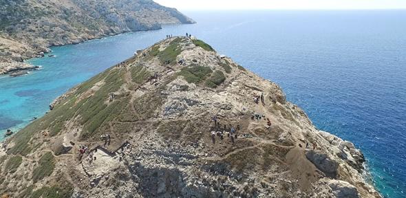
Until recently, the island of Keros, located in the Cyclades, south of Naxos, was known for ritual activities dating from 4,500 years ago involving broken marble figurines. Now new excavations are showing that the promontory of Dhaskalio (now a tiny islet because of sea level rise), at the west end of the island next to the sanctuary, was almost entirely covered by remarkable monumental constructions built using stone brought painstakingly from Naxos, some 10km distant.

Professor Colin Renfrew of the University of Cambridge, Co-Director of the excavation, suggested that the promontory, with its narrow causeway to the main island, “may have become a focus because it formed the best natural harbour on Keros, and had an excellent view of the north, south and west Aegean”.

The promontory was naturally shaped like a pyramid, and the skilled builders of Dhaskalio enhanced this shape by creating a series of massive terrace walls which made it look more like a stepped pyramid. On the flat surfaces formed by the terraces, the builders used stone imported from Naxos to construct impressive, gleaming structures.

The research team, led by archaeologists from the University of Cambridge, the Ephorate of the Cyclades and the Cyprus Institute, have calculated that more than 1000 tons of stone were imported, and that almost every possible space on the island was built on, giving the impression of a single large monument jutting out of the sea. The complex is the largest known in the Cyclades at the time.

Renfrew noted that “investigations at multiple points throughout the site have given unique insight into how the architecture was organised and how people moved about the built environment”.

While excavating an impressive staircase in the lower terraces, archaeologists began to see the technical sophistication of this civilisation 1000 years before the famous palaces of the Mycenaeans. Underneath the stairs and within the walls they discovered sophisticated systems of drainage, signalling that the architecture was multipurpose and carefully planned in advance. Tests are now underway to discover whether the drains were for managing clean water or sewage.

What was the reason for this massive undertaking here?

The rituals practised in the nearby sanctuary meant that this was already an important central place for the Cycladic islanders. Another aspect of the expansion of Dhaskalio is the use of new agricultural practices, whose study is led by Dr Evi Margaritis of the Cyprus Institute. She says: “Dhaskalio has already provided important evidence about the cultivation of olive and grape, two key new domesticates that expanded the horizons of agriculture in the third millennium. The environmental programme is revealing how agricultural strategies developed through the lifetime of the site.”

The excavated soil of the site is being examined in great detail for tiny clues in the form of burnt seeds, phytoliths (plant remnants preserved as silica), burnt wood, and animal and fish bones. Lipid and starch analysis on pottery and grinding stones is giving clues about food production and consumption.

Plant remains have been recovered in carbonised form, predominantly pulses and fruits such as grape, olives, figs and almonds, but also cereals such as emmer wheat and barley. Margaritis notes: “Keros was probably not self-sustaining, meaning that much of this food was imported: in the light of this evidence we need to reconsider what we know about existing networks to include food exchange”.

Another clue may be found in metalworking, the most important new technology of the third millennium BC. The inhabitants of Dhaskalio were proficient metalworkers, and the evidence for the associated technologies is strong everywhere on the site. No metal ore sources are located on Keros, so all raw materials were imported from elsewhere (other Cycladic islands such as Seriphos or Kythnos, or the mainland).

These imported ores were smelted just to the north of the sanctuary, where the winds were strongest, needed to achieve the very high temperatures required to extract metals from ores. Within the buildings of Dhaskalio, the melting of metals and casting of objects were commonplace.

The new excavations have found two metalworking workshops, full of metalworking debris and related objects. In one of these rooms a lead axe was found, with a mould used for making copper daggers, along with dozens of ceramic fragments (such as tuyères, the ceramic end of a bellows, used to force air into the fire to increase its temperature) covered in copper spills. In another room, which only appeared at the end of excavation this year, the top of an intact clay oven was found, indicating another metalworking area, which will be excavated next year.

What is the significance of the metalworking finds?

Dr Michael Boyd of the University of Cambridge, Co-Director of the excavation, says: “At a time when access to raw materials and skills was very limited, metalworking expertise seems to have been very much concentrated at Dhaskalio. What we are seeing here with the metalworking and in other ways is the beginnings of urbanisation: centralisation, meaning the drawing of far-flung communities into networks centred on the site, intensification in craft or agricultural production, aggrandisement in architecture, and the gradual subsuming of the ritual aspects of the sanctuary within the operation of the site. This gives us a clear insight into social change at Dhaskalio, from the earlier days where activities were centred on ritual practices in the sanctuary to the growing power of Dhaskalio itself in its middle years.”

The excavations on Keros are leading the charge of technical innovation in Aegean archaeology. All data are recorded digitally, using a new system called iDig – an app that runs on Apple’s iPads. For the first time in the Aegean, not only data from the excavation, but the results of study in the laboratory are all recorded in the same system, meaning that anyone on the excavation has access to all available data in real time. Three dimensional models are created at every stage in the digging process using a technique called photogrammetry; at the end of each season the trenches are recorded in detail by the Cyprus Institute’s laser scanning team.

The Cyprus Institute co-organised for a second year an educational programme during this year’s excavations with Cambridge University. Students from Greece, Australia, New Zealand, the USA, Canada, and the UK joined the excavation and gained valuable experience of up to the minute excavation and scientific techniques. The syllabus epitomised the twin goals of promoting science in archaeology and establishing the highest standards of teaching and research.

The project is organised under the auspices of the British School at Athens and conducted with permission of the Greek Ministry of Culture and Sport. The project is directed by Colin Renfrew and Michael Boyd of the McDonald Institute for Archaeological Research, University of Cambridge. The project is supported by the Institute for Aegean Prehistory, the Cyprus Institute, the McDonald Institute for Archaeological Research, the British Academy, the Society of Antiquaries of London, the Gerda Henkel Stiftung, National Geographic Society, Cosmote, Blue Star Lines, EZ-dot and private donors.


Creative Commons License
The text in this work is licensed under a Creative Commons Attribution 4.0 International License. For image use please see separate credits above.

AI ‘Scientist’ Finds That Toothpaste Ingredient May Help Fight Drug-Resistant Malaria

AI ‘scientist’ finds that toothpaste ingredient may help fight drug-resistant malaria


source: www.cam.ac.uk

An ingredient commonly found in toothpaste could be employed as an anti-malarial drug against strains of malaria parasite that have grown resistant to one of the currently-used drugs. This discovery, led by researchers at the University of Cambridge, was aided by Eve, an artificially-intelligent ‘robot scientist’.

Drug-resistant malaria is becoming an increasingly significant threat in Africa and south-east Asia, and our medicine chest of effective treatments is slowly depleting

Steve Oliver

When a mosquito infected with malaria parasites bites someone, it transfers the parasites into their bloodstream via its saliva. These parasites work their way into the liver, where they mature and reproduce. After a few days, the parasites leave the liver and hijack red blood cells, where they continue to multiply, spreading around the body and causing symptoms, including potentially life-threatening complications.

Malaria kills over half a million people each year, predominantly in Africa and south-east Asia. While a number of medicines are used to treat the disease, malaria parasites are growing increasingly resistant to these drugs, raising the spectre of untreatable malaria in the future.

Now, in a study published today in the journal Scientific Reports, a team of researchers employed the Robot Scientist ‘Eve’ in a high-throughput screen and discovered that triclosan, an ingredient found in many toothpastes, may help the fight against drug-resistance.

When used in toothpaste, triclosan prevents the build-up of plaque bacteria by inhibiting the action of an enzyme known as enoyl reductase (ENR), which is involved in the production of fatty acids.

Scientists have known for some time that triclosan also inhibits the growth in culture of the malaria parasite Plasmodium during the blood-stage, and assumed that this was because it was targeting ENR, which is found in the liver. However, subsequent work showed that improving triclosan’s ability to target ENR had no effect on parasite growth in the blood.

Working with ‘Eve’, the research team discovered that in fact, triclosan affects parasite growth by specifically inhibiting an entirely different enzyme of the malaria parasite, called DHFR. DHFR is the target of a well-established antimalarial drug, pyrimethamine; however, resistance to the drug among malaria parasites is common, particularly in Africa. The Cambridge team showed that triclosan was able to target and act on this enzyme even in pyrimethamine-resistant parasites.

“Drug-resistant malaria is becoming an increasingly significant threat in Africa and south-east Asia, and our medicine chest of effective treatments is slowly depleting,” says Professor Steve Oliver from the Cambridge Systems Biology Centre and the Department of Biochemistry at the University of Cambridge. “The search for new medicines is becoming increasingly urgent.”

Because triclosan inhibits both ENR and DHFR, the researchers say it may be possible to target the parasite at both the liver stage and the later blood stage.

Lead author Dr Elizabeth Bilsland, now an assistant professor at the University of Campinas, Brazil, adds: “The discovery by our robot ‘colleague’ Eve that triclosan is effective against malaria targets offers hope that we may be able to use it to develop a new drug. We know it is a safe compound, and its ability to target two points in the malaria parasite’s lifecycle means the parasite will find it difficult to evolve resistance.”

Robot scientist Eve was developed by a team of scientists at the Universities of Manchester, Aberystwyth, and Cambridge to automate – and hence speed up – the drug discovery process by automatically developing and testing hypotheses to explain observations, run experiments using laboratory robotics, interpret the results to amend their hypotheses, and then repeat the cycle, automating high-throughput hypothesis-led research.

Professor Ross King from the Manchester Institute of Biotechnology at the University of Manchester, who led the development of Eve, says: “Artificial intelligence and machine learning enables us to create automated scientists that do not just take a ‘brute force’ approach, but rather take an intelligent approach to science. This could greatly speed up the drug discovery progress and potentially reap huge rewards.”

The research was supported by the Biotechnology & Biological Sciences Research Council, the European Commission, the Gates Foundation and FAPESP (São Paulo Research Foundation).

Reference
Bilsland, E et al. Plasmodium dihydrofolate reductase is a second enzyme target for the antimalarial action of triclosan. Scientific Reports; 18 Jan 2018; DOI: 10.1038/s41598-018-19549-x


Creative Commons License
The text in this work is licensed under a Creative Commons Attribution 4.0 International License. For image use please see separate credits above.


How Incurable Mitochondrial Diseases Strike Previously Unaffected Families

How incurable mitochondrial diseases strike previously unaffected families

source: www.cam.ac.uk

Researchers have shown for the first time how children can inherit a severe – potentially fatal – mitochondrial disease from a healthy mother. The study, led by researchers from the MRC Mitochondrial Biology Unit at the University of Cambridge, reveals that healthy people harbour mutations in their mitochondrial DNA and explains how cases of severe mitochondrial disease can appear unexpectedly in previously unaffected families.

We know that these devastating mitochondrial mutations can pop up in families without any previous history, but previously we didn’t know how that happened. We were surprised to find that egg cells in healthy females all carry a few defects in their mitochondrial DNA

Patrick Chinnery

Mitochondrial diseases caused by mutations in mitochondrial DNA are rare, affecting approximately 1 in 10,000 births, but can cause severe conditions. For example, Leigh Syndrome is a severe brain disorder causing progressive loss of mental and movement abilities, which usually becomes apparent in the first year of life and typically results in death within two to three years.

Mitochondria are the powerhouses inside our cells, producing energy and carrying their own DNA instructions (separate from the DNA in the nucleus of every cell). Mitochondria are inherited from a person’s mother via the egg.

In the study, published in Nature Cell Biology, the researchers isolated mouse and human female embryonic germ cells – the cells that will go on to be egg cells in an adult woman – and tested their mitochondrial DNA.

They found that a variety of mutations were present in the mitochondrial DNA in the developing egg cells of all 12 of the human embryos studied, showing that low levels of mitochondrial DNA mutations are carried by healthy humans.

Professor Patrick Chinnery, from the MRC Mitochondrial Biology Unit and the Department of Clinical Neurosciences at the University of Cambridge, said: “We know that these devastating mitochondrial mutations can pop up in families without any previous history, but previously we didn’t know how that happened. We were surprised to find that egg cells in healthy females all carry a few defects in their mitochondrial DNA.”

For most of the human genome, mutations are kept in check by the processes of sexual reproduction, when eggs and sperm combine; however, mitochondria replicate asexually and mitochondrial DNA is inherited unchanged from the mother’s egg. This means that over time mutations can accumulate which, if left unchecked over generations, could eventually lead to malfunction and disease in offspring.

This conundrum led researchers to predict that a “bottleneck,” where only healthy mitochondria survive, may explain how mitochondria are kept healthy down the generations.

In this study, the researchers identified and measured this bottleneck for the first time in developing human egg cells. In these cells, the number of mitochondria decreased to approximately 100 mitochondria per cell, compared to around 100,000 mitochondria in a mature egg cell.

In a mature cell, a few faulty mitochondria could hide unnoticed amongst the thousands of healthy mitochondria, but the small number of mitochondria in the cell during the bottleneck means that the effects of faulty mitochondria are no longer masked.

The exact mechanism by which cells with unhealthy mitochondria are eliminated is not yet known, but since developing egg cells need a lot of energy – produced by the mitochondria – the researchers suggest that after the bottleneck stage, eggs cells containing damaged mitochondria cannot generate enough energy to mature and are lost.

This study found every developing egg cell may carry a few faulty mitochondria, so occasionally, by chance, after the bottleneck these could be the mitochondria that repopulate the egg cell. The scientists suggest that if the quality-control step fails, then this faulty egg could survive and develop into a child with a mitochondrial disease.

Professor Patrick Chinnery said: “Unfortunately, the purification process is not perfect, and occasionally defective mitochondria leak through. This can cause a severe disease in a child, despite no one else in the family having been affected.”

Mitochondrial diseases are currently incurable, although a new IVF technique of mitochondrial transfer gives families affected by mitochondrial disease the chance of having healthy children – removing affected mitochondria from an egg or embryo and replacing them with healthy ones from a donor.

The study authors also suggest that this process could be relevant for human aging. Professor Chinnery added: “Previously it was assumed that the mitochondrial DNA mutations that have been associated with diseases of ageing, such as Alzheimer’s disease, Parkinson’s disease and other neurodegenerative disorders, happened over a person’s lifetime. This study shows how some of these mutations can be inherited from your mother, potentially predisposing you to late onset brain diseases.”

Professor Chinnery is a Wellcome Trust Senior Research Fellow and the researchers were funded by Wellcome, the Medical Research Council and the National Institute for Health Research.

Dr Nathan Richardson, MRC Head of Molecular and Cellular Medicine, said: “This is an exciting study that reveals important new insights into how mitochondrial diseases develop and are inherited between generations. The researchers have made great use of the tissues available from the MRC-Wellcome Human Developmental Biology Resource (HDBR). The HDBR is an internationally unique biobank resource that provides human embryonic and foetal tissue, donated through elective terminations, facilitating research into a large number of distressing medical disorders, such as mitochondrial diseases.”

Reference
Floros, V et al. Segregation of mitochondrial DNA heteroplasmy through a developmental genetic bottleneck in human embryos. Nature Cell Biology; 15 Jan 2018; DOI: 10.1038/41556-017-0017-8


Creative Commons License
The text in this work is licensed under a Creative Commons Attribution 4.0 International License. For image use please see separate credits above.

Astronomers Detect ‘Whirlpool’ Movement In Earliest Galaxies

Astronomers detect ‘whirlpool’ movement in earliest galaxies

source: www.cam.ac.uk

Astronomers have looked back to a time soon after the Big Bang, and have discovered swirling gas in some of the earliest galaxies to have formed in the Universe. These ‘newborns’ – observed as they appeared nearly 13 billion years ago – spun like a whirlpool, similar to our own Milky Way. This is the first time that it has been possible to detect movement in galaxies at such an early point in the Universe’s history.

We’ve never been able to see the formation of galaxies in such detail, and we’ve never been able to measure the movement of gas in galaxies so early in the Universe’s history.

Stefano Carniani

An international team led by Dr Renske Smit from the Kavli Institute of Cosmology at the University of Cambridge used the Atacama Large Millimeter/submillimeter Array (ALMA) in Chile to open a new window onto the distant Universe, and have for the first time been able to identify normal star-forming galaxies at a very early stage in cosmic history with this telescope. The results are reported in the journal Nature, and will be presented at the 231st meeting of the American Astronomical Society.

Light from distant objects takes time to reach Earth, so observing objects that are billions of light years away enables us to look back in time and directly observe the formation of the earliest galaxies. The Universe at that time, however, was filled with an obscuring ‘haze’ of neutral hydrogen gas, which makes it difficult to see the formation of the very first galaxies with optical telescopes.

Smit and her colleagues used ALMA to observe two small newborn galaxies, as they existed just 800 million years after the Big Bang. By analysing the spectral ‘fingerprint’ of the far-infrared light collected by ALMA, they were able to establish the distance to the galaxies and, for the first time, see the internal motion of the gas that fuelled their growth.

“Until ALMA, we’ve never been able to see the formation of galaxies in such detail, and we’ve never been able to measure the movement of gas in galaxies so early in the Universe’s history,” said co-author Dr Stefano Carniani, from Cambridge’s Cavendish Laboratory and Kavli Institute of Cosmology.

The researchers found that the gas in these newborn galaxies swirled and rotated in a whirlpool motion, similar to our own galaxy and other, more mature galaxies much later in the Universe’s history. Despite their relatively small size – about five times smaller than the Milky Way – these galaxies were forming stars at a higher rate than other young galaxies, but the researchers were surprised to discover that the galaxies were not as chaotic as expected.

“In the early Universe, gravity caused gas to flow rapidly into the galaxies, stirring them up and forming lots of new stars – violent supernova explosions from these stars also made the gas turbulent,” said Smit, who is a Rubicon Fellow at Cambridge, sponsored by the Netherlands Organisation for Scientific Research. “We expected that young galaxies would be dynamically ‘messy’, due to the havoc caused by exploding young stars, but these mini-galaxies show the ability to retain order and appear well regulated. Despite their small size, they are already rapidly growing to become one of the ‘adult’ galaxies like we live in today.”

The data from this project on small galaxies paves the way for larger studies of galaxies during the first billion years of cosmic time. The research was funded in part by the European Research Council and the UK Science and Technology Facilities Council (STFC).

Reference:
Renske Smit et al. ‘Rotation in [C II]-emitting gas in two galaxies at a redshift of 6.8.’ Nature (2018). DOI: 10.1038/nature24631


Researcher profile: Renske Smit

Dr Renske Smit is a postdoctoral researcher and Rubicon Fellow at the Kavli Institute of Cosmology and is supported by the Netherlands Organisation for Scientific Research. Prior to arriving in Cambridge in 2016, she was a postdoctoral researcher at Durham University and a PhD student at Leiden University in the Netherlands.

Her research aims to understand how the first sources of light in the Universe came to be. In her daily work, she studies images of deep space, taken by telescopes such as the Hubble Space Telescope. To gather data, she sometimes travels to places such as Chile or Hawaii to work on big telescopes.

“In Cambridge, I have joined a team working on the James Webb Space Telescope, the most ambitious and expensive telescope ever built,” she says. “With this telescope, we might be able to see the very first stars for the first time. To have this kind of privileged access to world-leading data is truly a dream come true.

“I would like to contribute to changing the perception of what a science professor looks like. Women in the UK and worldwide are terribly underrepresented in science and engineering and as a result, people may feel women either don’t have the inclination or the talent to do science. I hope that one day I will teach students that don’t feel they represent the professor stereotype and make them believe in their own talent.”


Creative Commons License
The text in this work is licensed under a Creative Commons Attribution 4.0 International License. For image use please see separate credits above.

Harnessing the Power of Algae: New, Greener Fuel Cells Move Step Closer To Reality

Harnessing the power of algae: new, greener fuel cells move step closer to reality

source: www.cam.ac.uk

A new design of algae-powered fuel cells that is five times more efficient than existing plant and algal models, as well as being potentially more cost-effective to produce and practical to use, has been developed by researchers at the University of Cambridge.

This a big step forward in the search for alternative, greener fuels

Paolo Bombelli

As the global population increases, so too does energy demand. The threat of climate change means that there is an urgent need to find cleaner, renewable alternatives to fossil fuels that do not contribute extensive amounts of greenhouse gases with potentially devastating consequences on our ecosystem. Solar power is considered to be a particularly attractive source as on average the Earth receives around 10,000 times more energy from the sun in a given time than is required by human consumption.

In recent years, in addition to synthetic photovoltaic devices, biophotovoltaics (BPVs, also known as biological solar-cells) have emerged as an environmentally-friendly and low-cost approach to harvesting solar energy and converting it into electrical current. These solar cells utilise the photosynthetic properties of microorganisms such as algae to convert light into electric current that can be used to provide electricity.

During photosynthesis, algae produce electrons, some of which are exported outside the cell where they can provide electric current to power devices. To date, all the BPVs demonstrated have located charging (light harvesting and electron generation) and power delivery (transfer to the electrical circuit) in a single compartment; the electrons generate current as soon as they have been secreted.

In a new technique described in the journal Nature Energy, researchers from the departments of Biochemistry, Chemistry and Physics have collaborated to develop a two-chamber BPV system where the two core processes involved in the operation of a solar cell – generation of electrons and their conversion to power – are separated.

“Charging and power delivery often have conflicting requirements,” explains Kadi Liis Saar, of the Department of Chemistry. “For example, the charging unit needs to be exposed to sunlight to allow efficient charging, whereas the power delivery part does not require exposure to light but should be effective at converting the electrons to current with minimal losses.”

Building a two-chamber system allowed the researchers to design the two units independently and through this optimise the performance of the processes simultaneously.

“Separating out charging and power delivery meant we were able to enhance the performance of the power delivery unit through miniaturisation,” explains Professor Tuomas Knowles from the Department of Chemistry and the Cavendish Laboratory. “At miniature scales, fluids behave very differently, enabling us to design cells that are more efficient, with lower internal resistance and decreased electrical losses.”

The team used algae that had been genetically modified to carry mutations that enable the cells to minimise the amount of electric charge dissipated non-productively during photosynthesis. Together with the new design, this enabled the researchers to build a biophotovoltaic cell with a power density of 0.5 W/m2, five times that of their previous design. While this is still only around a tenth of the power density provided by conventional solar fuel cells, these new BPVs have several attractive features, they say.

“While conventional silicon-based solar cells are more efficient than algae-powered cells in the fraction of the sun’s energy they turn to electrical energy, there are attractive possibilities with other types of materials,” says Professor Christopher Howe from the Department of Biochemistry. “In particular, because algae grow and divide naturally, systems based on them may require less energy investment and can be produced in a decentralised fashion.”

Separating the energy generation and storage components has other advantages, too, say the researchers. The charge can be stored, rather than having to be used immediately – meaning that the charge could be generated during daylight and then used at night-time.

While algae-powered fuel cells are unlikely to generate enough electricity to power a grid system, they may be particularly useful in areas such as rural Africa, where sunlight is in abundance but there is no existing electric grid system. In addition, whereas semiconductor-based synthetic photovoltaics are usually produced in dedicated facilities away from where they are used, the production of BPVs could be carried out directly by the local community, say the researchers.

“This a big step forward in the search for alternative, greener fuels,” says Dr Paolo Bombelli, from the Department of Biochemistry. “We believe these developments will bring algal-based systems closer to practical implementation.”

The research was supported by the Leverhulme Trust, the Engineering and Physical Sciences Research Council and the European Research Council.

Reference
Saar, KL et al. Enhancing power density of biophotovoltaics by decoupling storage and power delivery. Nature Energy; 9 Jan 2018; DOI: 10.1038/s41560-017-0073-0


Researcher Profile: Dr Paolo Bombelli

Dr Paolo Bombelli is a post-doctoral researcher in the Department of Biochemistry, where his research looks to utilise the photosynthetic and metabolic activity of plants, algae and bacteria to create biophotovoltaic devices, a sustainable source of renewable current. He describes himself as “a plants, algae and bacteria electrician”.

“Photosynthesis generates a flow of electrons that keeps plants, algae and other photosynthetic organisms alive,” he explains. “These electrons flow though biological wires and, like the electrical current obtained from a battery and used to power a radio, they are the driving force for any cellular activity.”

Dr Bombelli’s fascination with this area of research began during his undergraduate studies at the University of Milan.

“Plants, algae and photosynthetic bacteria are the oldest, most common and effective solar panels on our planet,” he says. “For billions of years they have been harnessing the energy of the sun and using it to provide oxygen, food and materials to support life. With my work I aim to provide new ways to embrace the potential of these fantastic photosynthetic organisms.”

His work is highly cross-disciplinary, with input from the Departments of Biochemistry, Plant Sciences, Chemistry and Physics, and the Institute for Manufacturing, as well as from researchers at Imperial College London, UCL, the University of Brighton, the Institute for Advanced Architecture of Catalonia in Spain and the University of Cape Town, South Africa.

“Universities are great places to work and so they attract many people,” he says. “People choose to come to Cambridge because they know the ideas they generate here will go on to change the world.”

In 2016, Dr Bombelli won a Public Engagement with Research Award by the University of Cambridge for his work engaging audiences at more than 40 public events, including science festivals and design fairs, reaching thousands of people in seven countries. His outreach work included working with Professor Chris Howe to develop a prototype ‘green bus shelter’ where plants, classical solar panels and bio-electrochemical systems operate in synergy in a single structure.


Creative Commons License
The text in this work is licensed under a Creative Commons Attribution 4.0 International License. For image use please see separate credits above.