Over half a million people take part in largest ever study of psychological sex differences and autistic traits

Scientists at the University of Cambridge have completed the world’s largest ever study of typical sex differences and autistic traits. They tested and confirmed two long-standing psychological theories: the Empathising-Systemising theory of sex differences and the Extreme Male Brain theory of autism.
Big data is important to draw conclusions that are replicable and robust. This is an example of how scientists can work with the media to achieve big data science
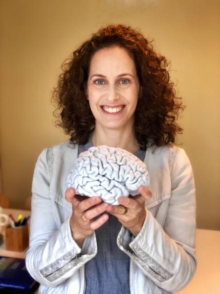
David Greenberg
Working with the television production company Channel 4, they tested over half a million people, including over 36,000 autistic people. The results are published today in the Proceedings of the National Academy of Sciences.
The Empathising-Systemising theory predicts that women, on average, will score higher than men on tests of empathy, the ability to recognize what another person is thinking or feeling, and to respond to their state of mind with an appropriate emotion. Similarly, it predicts that men, on average, will score higher on tests of systemising, the drive to analyse or build rule-based systems.
The Extreme Male Brain theory predicts that autistic people, on average, will show a masculinised shift on these two dimensions: namely, that they will score lower than the typical population on tests of empathy and will score the same as if not higher than the typical population on tests of systemising.
Whereas both theories have been confirmed in previous studies of relatively modest samples, the new findings come from a massive sample of 671,606 people, which included 36,648 autistic people. They were replicated in a second sample of 14,354 people. In this new study, the scientists used very brief 10-item measures of empathy, systemising, and autistic traits.
Using these short measures, the team identified that in the typical population, women, on average, scored higher than men on empathy, and men, on average, scored higher than women on systemising and autistic traits. These sex differences were reduced in autistic people. On all these measures, autistic people’s scores, on average, were ‘masculinised’: that is, they had higher scores on systemising and autistic traits and lower scores on empathy, compared to the typical population.
The team also calculated the difference (or ‘d-score’) between each individual’s score on the systemising and empathy tests. A high d-score means a person’s systemising is higher than their empathy, and a low d-score means their empathy is higher than their systemising.
They found that in the typical population, men, on average, had a shift towards a high d-score, whereas women, on average, had a shift towards a low d-score. Autistic individuals, on average, had a shift towards an even higher d-score than typical males. Strikingly, d-scores accounted for 19 times more of the variance in autistic traits than other variables, including sex.
Finally, men, on average, had higher autistic trait scores than women. Those working in STEM (Science, Technology, Engineering and Mathematics), on average, had higher systemising and autistic traits scores than those in non-STEM occupations. And conversely, those working in non-STEM occupations, on average, had had higher empathy scores than those working in STEM.
In the paper, the authors discuss how it is important to bear in mind that differences observed in this study apply only to group averages, not to individuals. They underline that these data say nothing about an individual based on their gender, autism diagnosis, or occupation. To do that would constitute stereotyping and discrimination, which the authors strongly oppose.
Further, the authors reiterate that the two theories are applicable to only two dimensions of typical sex differences: empathy and systemising. They do not apply to all sex differences, such as aggression, and to extrapolate the theories beyond these two dimensions would be a misinterpretation.
Finally, the authors highlight that although autistic people on average struggle with ‘cognitive’ empathy – recognizing other people’s thoughts and feelings – they nevertheless have intact ‘affective’ empathy – they care about others. It is a common misunderstanding that autistic people struggle with all forms of empathy, which is untrue.
Dr Varun Warrier, from the Cambridge team, said: “These sex differences in the typical population are very clear. We know from related studies that individual differences in empathy and systemising are partly genetic, partly influenced by our prenatal hormonal exposure, and partly due to environmental experience. We need to investigate the extent to which these observed sex differences are due to each of these factors, and how these interact.”
Dr David Greenberg, from the Cambridge team, said: “Big data is important to draw conclusions that are replicable and robust. This is an example of how scientists can work with the media to achieve big data science.”
Dr Carrie Allison, from the Cambridge team, said: “We are grateful to both the general public and to the autism community for participating in this research. The next step must be to consider the relevance of these findings for education, and support where needed.”
Professor Simon Baron-Cohen, Director of the Autism Research Centre at Cambridge who proposed these two theories nearly two decades ago, said: “This research provides strong support for both theories. This study also pinpoints some of the qualities autistic people bring to neurodiversity. They are, on average, strong systemisers, meaning they have excellent pattern-recognition skills, excellent attention to detail, and an aptitude in understanding how things work. We must support their talents so they achieve their potential – and society benefits too.”
This study was supported by the Autism Research Trust, the Medical Research Council, Wellcome, and the Templeton World Charity Foundation., Inc. It was conducted in association with the NIHR CLAHRC for Cambridgeshire and Peterborough NHS Foundation Trust, and the NIHR Cambridge Biomedical Research Centre.
Reference
Greenberg, DM et al. Testing the Empathizing-Systemising theory of sex differences and the Extreme Male Brain theory of autism in half a million people. PNAS; 12 Nov 2018; DOI: 10.1073/pnas.1811032115
If you’d like to complete these measures and participate in studies at the Autism Research Centre please register here.

The text in this work is licensed under a Creative Commons Attribution 4.0 International License. Images, including our videos, are Copyright ©University of Cambridge and licensors/contributors as identified. All rights reserved. We make our image and video content available in a number of ways – as here, on our main website under its Terms and conditions, and on a range of channels including social media that permit your use and sharing of our content under their respective Terms.


. © The British Library Board")