Sir Isaac Newton’s Cambridge papers added to UNESCO’s Memory of the World Register
The Cambridge papers of Sir Isaac Newton, including early drafts and Newton’s annotated copies of Principia Mathematica – a work that changed the history of science – have been added to UNESCO’s International Memory of the World Register.
Newton’s papers are among the world’s most important collections in the western scientific tradition and are one of the Library’s most treasured collections.
Katrina Dean
Held at Cambridge University Library, Newton’s scientific and mathematical papers represent one of the most important archives of scientific and intellectual work on universal phenomena. They document the development of his thought on gravity, calculus and optics, and reveal ideas worked out through painstaking experiments, calculations, correspondence and revisions.
In combination with alchemical papers at King’s College, Cambridge and his notebooks and correspondence at Trinity College, Cambridge and the Fitzwilliam Museum, this represents the largest and most important collection of Newton’s papers worldwide.
Katrina Dean, Curator of Scientific Collections at Cambridge University Library said: “Newton’s papers are among the world’s most important collections in the western scientific tradition and are one of the Library’s most treasured collections. They were the first items to be digitised and added to the Cambridge Digital Library in 2011 and featured in our 600th anniversary exhibition Lines of Thought last year. In 2017, their addition to the UNESCO International Memory of the World Register recognises their unquestionable international importance.”
The Memory of the World Project is an international initiative to safeguard the documentary heritage of humanity against collective amnesia, neglect, the ravages of time and climatic conditions, and wilful and deliberate destruction. It calls for the preservation of valuable archival, library and private collections all over the world, as well as the reconstitution of dispersed or displaced documentary heritage, and the increased accessibility to and dissemination of these items.
Newton’s Cambridge papers, and those at the Royal Society, now join the archive of Winston Churchill, held at Cambridge University’s Churchill Archives Centre, on the UNESCO Register. They also join Newton’s theological and alchemical papers at the National Library of Israel, which were added in 2015.
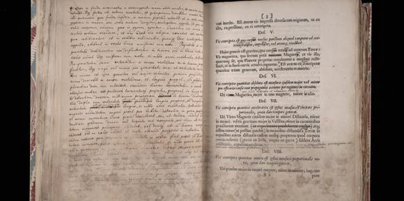
The chief attractions in the Cambridge collection are Newton’s own copies of the first edition of the Principia (1687), covered with his corrections, revisions and additions for the second edition.

The Cambridge papers also include significant correspondence with natural philosophers and mathematicians including Henry Oldenberg, Secretary of the Royal Society, Edmond Halley, the Astronomer Royal who persuaded Newton to publish Principia, Richard Bentley, the Master of Trinity College, and John Collins, mathematician and fellow of the Royal Society who became an important collector of Newton’s works.
Added Dean: “One striking illustration of Newton’s experimental approach is in his ‘Laboratory Notebook’, which includes details of his investigations into light and optics in order to understand the nature of colour. His essay ‘Of Colours’ includes a diagram that illustrates the experiment in which he inserted a bodkin into his eye socket to put pressure on the eyeball to try to replicate the sensation of colour in normal sight.”
Another important item is Newton’s so-called ‘Waste Book’, a large notebook inherited from his stepfather. From 1664, he used the blank pages for optical and mathematical calculations and gradually mastered the analysis of curved lines, surfaces and solids. By 1665, he had invented the method of calculus. Newton later used the dated, documentary evidence provided by the Waste Book to argue his case in the priority dispute with Gottfried Wilhelm Leibniz over the invention of the calculus.
Cambridge University Librarian Jess Gardner said: “Newton’s work and life continue to attract wonder and new perspectives on our place in the Universe. Cambridge University Library will continue to work with scholars and curators worldwide to make Newton’s papers accessible now and for future generations.”
Isaac Newton entered Trinity College as an undergraduate in 1661 and became a Fellow in 1667. In 1669, he became Lucasian Professor of Mathematics at Cambridge University, a position he held until 1701.
Among the more personal items in the Cambridge collections are Newton’s daily concerns as recorded in an undergraduate notebook which records Newton’s expenditure on white wine, wafers, shoe-strings and ‘a paire of stockings’, along with a guide to Latin pronunciation.
A notebook of 1662-1669 records Newton’s sins before and after Whitsunday of 1662, written in a coded shorthand and first deciphered between 1872 and 1888. Among them are ‘Eating an apple at Thy house’, ‘Robbing my mothers box of plums and sugar’ along with the more serious ‘Wishing death and hoping it to some’ before a list of his expenses. These included chemicals, two furnaces and a recent edition of one of the most comprehensive compilations of alchemical writings in the western tradition Theatrum chemicum, edited by the publisher Lazarus Zetzner.
Cambridge University Library is also hosting a series of talks open to the public by Sarah Dry and Patricia Fara on Newton’s manuscripts and Newton’s role in Enlightenment culture and polite society on December 7 and December 14 respectively. For details and bookings, see: http://www.lib.cam.ac.uk/using-library/whats

The text in this work is licensed under a Creative Commons Attribution 4.0 International License. For image use please see separate credits above.