LIGO detects gravitational waves for third time

Results confirm new population of black holes.
Each new detection enables us to explore new phenomena of these mysterious and fascinating objects.
Ulrich Sperhake
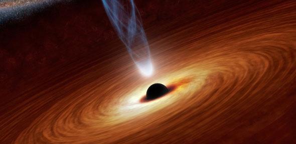
The Laser Interferometer Gravitational-wave Observatory (LIGO) has made a third detection of gravitational waves, ripples in space and time, demonstrating that a new window in astronomy has been firmly opened. As was the case with the first two detections, the waves were generated when two black holes collided to form a larger black hole.
The newfound black hole formed by the merger has a mass about 49 times that of our sun. “With this third confirmed detection we are uncovering the population of black holes in the Universe for the first time,” said Christopher Moore from the University of Cambridge’s Department of Applied Mathematics and Theoretical Physics (DAMTP), who is part of the LIGO Scientific Collaboration.
The new detection occurred during LIGO’s current observing run, which began November 30, 2016, and will continue through the summer. LIGO is an international collaboration with members around the globe. Its observations are carried out by twin detectors—one in Hanford, Washington, and the other in Livingston, Louisiana—operated by Caltech and MIT with funding from the United States National Science Foundation (NSF).
The LIGO group in Cambridge consists of seven researchers spread across DAMTP, the Cavendish Laboratory and the Institute of Astronomy.
“Answering key questions about the formation history of astrophysical black holes and their role in the evolution of the universe critically relies on applying a statistical analysis to a sufficiently large sample of observations,” said Dr Ulrich Sperhake, head of the group in DAMTP. “Each new detection not only strengthens our confidence in the theoretical modelling, but enables us to explore new phenomena of these mysterious and fascinating objects.”
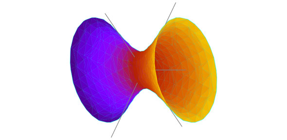
One of the interests of the Cambridge group is testing Einstein’s theory of general relativity. “This particular source of gravitational waves is the furthest detected so far. This allows us to test our understanding of the propagation of gravitational waves across cosmological distances, by means of which we constrained any signs of wave dispersion to unprecedented precision,” said Dr Michalis Agathos, a postdoctoral researcher at DAMTP.
The LIGO-Virgo team is continuing to search the latest LIGO data for signs of space-time ripples from the far reaches of the cosmos. They are also working on technical upgrades for LIGO’s next run, scheduled to begin in late 2018, during which the detectors’ sensitivity will be further improved.
“With the third confirmed detection of gravitational waves from the collision of two black holes, LIGO is establishing itself as a powerful observatory for revealing the dark side of the universe,” said David Reitze of Caltech, executive director of the LIGO Laboratory. “While LIGO is uniquely suited to observing these types of events, we hope to see other types of astrophysical events soon, such as the violent collision of two neutron stars.”
LIGO is funded by the National Science Foundation (NSF), and operated by MIT and Caltech, which conceived and built the project. Financial support for the Advanced LIGO project was led by NSF with Germany (Max Planck Society), the UK (Science and Technology Facilities Council) and Australia (Australian Research Council) making significant commitments and contributions to the project. More than 1,000 scientists from around the world participate in the effort through the LIGO Scientific Collaboration, which includes the GEO Collaboration. LIGO partners with the Virgo Collaboration, a consortium including 280 additional scientists throughout Europe supported by the Centre National de la Recherche Scientifique (CNRS), the Istituto Nazionale di Fisica Nucleare (INFN), and Nikhef, as well as Virgo’s host institution, the European Gravitational Observatory. Additional partners are listed at: http://ligo.org/partners.php.

The text in this work is licensed under a Creative Commons Attribution 4.0 International License. For image use please see separate credits above.