Astronomers detect ‘whirlpool’ movement in earliest galaxies

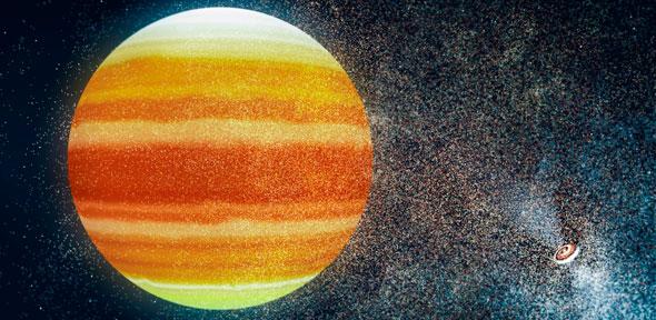
Astronomers have looked back to a time soon after the Big Bang, and have discovered swirling gas in some of the earliest galaxies to have formed in the Universe. These ‘newborns’ – observed as they appeared nearly 13 billion years ago – spun like a whirlpool, similar to our own Milky Way. This is the first time that it has been possible to detect movement in galaxies at such an early point in the Universe’s history.
We’ve never been able to see the formation of galaxies in such detail, and we’ve never been able to measure the movement of gas in galaxies so early in the Universe’s history.
Stefano Carniani
An international team led by Dr Renske Smit from the Kavli Institute of Cosmology at the University of Cambridge used the Atacama Large Millimeter/submillimeter Array (ALMA) in Chile to open a new window onto the distant Universe, and have for the first time been able to identify normal star-forming galaxies at a very early stage in cosmic history with this telescope. The results are reported in the journal Nature, and will be presented at the 231st meeting of the American Astronomical Society.
Light from distant objects takes time to reach Earth, so observing objects that are billions of light years away enables us to look back in time and directly observe the formation of the earliest galaxies. The Universe at that time, however, was filled with an obscuring ‘haze’ of neutral hydrogen gas, which makes it difficult to see the formation of the very first galaxies with optical telescopes.
Smit and her colleagues used ALMA to observe two small newborn galaxies, as they existed just 800 million years after the Big Bang. By analysing the spectral ‘fingerprint’ of the far-infrared light collected by ALMA, they were able to establish the distance to the galaxies and, for the first time, see the internal motion of the gas that fuelled their growth.
“Until ALMA, we’ve never been able to see the formation of galaxies in such detail, and we’ve never been able to measure the movement of gas in galaxies so early in the Universe’s history,” said co-author Dr Stefano Carniani, from Cambridge’s Cavendish Laboratory and Kavli Institute of Cosmology.
The researchers found that the gas in these newborn galaxies swirled and rotated in a whirlpool motion, similar to our own galaxy and other, more mature galaxies much later in the Universe’s history. Despite their relatively small size – about five times smaller than the Milky Way – these galaxies were forming stars at a higher rate than other young galaxies, but the researchers were surprised to discover that the galaxies were not as chaotic as expected.
“In the early Universe, gravity caused gas to flow rapidly into the galaxies, stirring them up and forming lots of new stars – violent supernova explosions from these stars also made the gas turbulent,” said Smit, who is a Rubicon Fellow at Cambridge, sponsored by the Netherlands Organisation for Scientific Research. “We expected that young galaxies would be dynamically ‘messy’, due to the havoc caused by exploding young stars, but these mini-galaxies show the ability to retain order and appear well regulated. Despite their small size, they are already rapidly growing to become one of the ‘adult’ galaxies like we live in today.”
The data from this project on small galaxies paves the way for larger studies of galaxies during the first billion years of cosmic time. The research was funded in part by the European Research Council and the UK Science and Technology Facilities Council (STFC).
Reference:
Renske Smit et al. ‘Rotation in [C II]-emitting gas in two galaxies at a redshift of 6.8.’ Nature (2018). DOI: 10.1038/nature24631
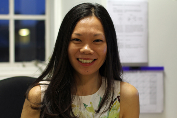
 Dr Renske Smit is a postdoctoral researcher and Rubicon Fellow at the Kavli Institute of Cosmology and is supported by the Netherlands Organisation for Scientific Research. Prior to arriving in Cambridge in 2016, she was a postdoctoral researcher at Durham University and a PhD student at Leiden University in the Netherlands.
Her research aims to understand how the first sources of light in the Universe came to be. In her daily work, she studies images of deep space, taken by telescopes such as the Hubble Space Telescope. To gather data, she sometimes travels to places such as Chile or Hawaii to work on big telescopes.
“In Cambridge, I have joined a team working on the James Webb Space Telescope, the most ambitious and expensive telescope ever built,” she says. “With this telescope, we might be able to see the very first stars for the first time. To have this kind of privileged access to world-leading data is truly a dream come true.
“I would like to contribute to changing the perception of what a science professor looks like. Women in the UK and worldwide are terribly underrepresented in science and engineering and as a result, people may feel women either don’t have the inclination or the talent to do science. I hope that one day I will teach students that don’t feel they represent the professor stereotype and make them believe in their own talent.”

The text in this work is licensed under a Creative Commons Attribution 4.0 International License. For image use please see separate credits above.


 Jakob Seidlitz is at PhD student on the NIH Oxford-Cambridge Scholars Programme. A graduate of the University of Rochester, USA, he spends half of his time in Cambridge and half at the National Institutes of Health in the USA.


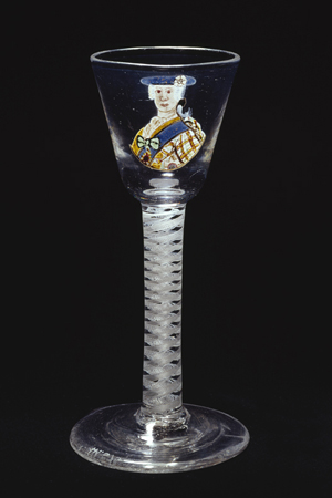 Two changes in the 20th century likely contributed further to increased glass sizes. Wine glasses started to be tailored in both shape and size for different wine varieties, both reflecting and contributing to a burgeoning market for wine appreciation, with larger glasses considered important in such appreciation. From 1990 onwards, demand for larger wine glasses by the US market was met by an increase in the size of glasses manufactured in England, where a ready market was also found.